• Pharmacogenomics studies how our genome influences medication responses, affecting both drug effectiveness and the likelihood of adverse reactions  (Gurwitz & Motulsky, 2007). Observing occurrences of adverse drug reactions (ADRs) at different rates in diverse populations can provide evidence for a genetic component to variability. These observations often originate from differences in the frequencies of specific gene alleles involved in drug metabolism or action pathways (Gurwitz & Motulsky, 2007). An identified challenge in applying pharmacogenomics at a clinical level is that much of the foundational research identifying relevant genetic variants was conducted primarily using populations of European ancestry. This narrow focus can create disparities, limiting the effectiveness and equity of pharmacogenomic testing when applied to individuals from other ancestral backgrounds (Lu, Lewis, & Traylor, 2017).

    The field of pharmacogenetics was conceptualized decades ago, yet its routine integration into clinical practice to guide prescribing medications has progressed somewhat slowly. ADRs are a problem in healthcare, revealing a continuing need for strategies to improve medication safety (Gurwitz & Motulsky, 2007). While some ADRs can be explained by variants in single genes, many drug responses likely arise from complex interactions involving multiple genes plus environmental influences. Early observations regarding population differences in drug responses stressed the importance of the genetic component. Applying pharmacogenetic insights more broadly in patient care promises to enhance drug safety and effectiveness (Gurwitz & Motulsky, 2007).

    The rise of Direct-to-Consumer (DTC) genetic testing companies, like 23andMe (RIP), now provides individuals direct access to some pharmacogenetic information, circumventing traditional healthcare intermediaries (Lu, Lewis, & Traylor, 2017). The study by Lu, Lewis & Taylor in 2017 evaluated DTC pharmacogenetic tests offered in the UK market, comparing them against established clinical guidelines and covering tests related to drug responses (Lu, Lewis, & Traylor, 2017). A principal finding was that the clinical usefulness and validity of these DTC tests varied substantially, with their applicability often strongly dependent on the user’s ancestral background because allele frequencies and local linkage disequilibrium (LD) patterns differ significantly across global populations (Lu, Lewis, & Traylor, 2017).

    The population differences have direct clinical relevance. For instance, CYP2C19 poor metabolizer alleles are more common in East Asian populations (~14%) than in European (~2%) or African (~4%) populations (Lu, Lewis, & Traylor, 2017). These findings imply standard clopidogrel doses may fail more often in East Asians, which emphasizes the need for population-aware guidance. Another example involves testing for abacavir hypersensitivity risk associated with HLA-B57:01*. Some tests use a tagging SNP that works well in Europeans but is uninformative in Africans and has variable accuracy in Asians due to differing LD patterns, necessitating direct HLA-B57:01* testing or population-specific tags (Lu, Lewis, & Traylor, 2017). Similarly, common variants in CYP2C9 and VKORC1 that influence warfarin dosing show different frequencies across populations suggesting dosing algorithms developed mainly in Europeans may require adjustments for other groups (Lu, Lewis, & Traylor, 2017).

    The findings reported by the Lu study emphasize the need for pharmacogenomic research to include diverse global populations to ensure findings are broadly applicable. Relying on genetic markers identified primarily in one ancestral group can lead to tests lacking accuracy or relevance for others (Lu, Lewis, & Traylor, 2017). Establishing comprehensive population-specific reference data and ensuring test reports and guidelines incorporate information about population-specific performance are needed for accurate clinical interpretation worldwide. While DTC testing can raise awareness, navigating these intricacies often benefits from guidance from knowledgeable healthcare professionals (Lu, Lewis, & Traylor, 2017). Addressing population diversity issues is important for the equitable use of pharmacogenomics to improve medication outcomes for everyone (Gurwitz & Motulsky, 2007).

    The frequencies of alleles influencing drug response vary among different human populations. Research focusing on European ancestry groups has led to potential gaps and inequities when applying genomic information across diverse populations (Lu, Lewis, & Traylor, 2017). Realizing the full potential of pharmacogenomics responsibly and equitably requires continued research in diverse populations, refining testing strategies, and developing ancestry-aware clinical resources (Gurwitz & Motulsky, 2007).

    References

    Gurwitz, D., & Motulsky, A. (2007). ‘Drug Reactions, Enzymes, and Biochemical Genetics‘: 50 years Later. Pharmacogenomics, 8(11), 1479-1484; https://doi.org/10.2217/14622416.8.11.1479.

    Lu, M., Lewis, C., & Traylor, M. (2017). Pharmacogenetic testing through the direct-to-consumer genetic testing company 23andMe. BMC Medical Genomics, 10(47). , https://doi.org/10.1186/s12920-017-0283-0.

  • Rett syndrome is a rare disorder with a classic presentation, as well as several variants with differing symptoms. It is generally caused by a random mutation in the MECP2 gene and causes a developmental regression in children, in which they begin to lose fine motor skills and other acquired skills within the first 7-18 months of life. Those affected by Rett syndrome develop normally up until the onset of symptoms. Symptoms can be severe but generally progress and the children could begin to develop autistic behaviors as well as seizures. Rett syndrome rarely affects males, as it is an X-linked gene. Symptoms have a range and can be very mild, or severe. Symptoms include loss of control over voluntary movements (walking, crawling), compulsive hand movements (clapping, washing), difficulty swallowing, and microcephaly (Neul & Eskind, 2023).

    Rett syndrome occurs in various stages with standard progression. After initial onset of symptoms, affected children go through a stage of rapid deterioration from the age of about 1 to 4 in which their symptoms progress over short periods of weeks or months. The next stage is characterized by a plateau of symptoms, sometimes with a slight improvement in speech and motor skills. In later stages, children with Rett syndrome slowly lose their mobility but can maintain most of their cognitive ability.

    The exact cause of Rett syndrome is unknown, but it is generally caused by random mutations in the MECP2 gene, which may lead to protein production problems in the developing brains of those with the mutations. Males with Rett syndrome typically die before infancy, due to the male chromosomal arrangement of XY. There are no known risk factors, but there may be a higher risk in families with a history of Rett syndrome, although this is not confirmed by any study (Mayo Clinic, 2022).

    Using the tool, NeuroTri2-VISDOT, to study the MECP2 gene, it is readily apparent that the MECP2 gene is highly expressive in the Cajal-Retzius (CR) cells at 18 weeks (Fig. 1; Fig. 2; Fig. 3). CR cells are responsible for organization of neurons in the fetal brain. This allows neurons to be sent to various layers of brain tissue. CR cells are also known to attract early serotonin input, which may allow for synaptic contact in neurons. Reelin is a protein secreted by CR cells which allows CR cells to control the detachment of radial glia and the organization of the layers of the cortex.

    CR cells in Alzheimer’s patients are diminished in number and have decreased synaptic contact with adjacent cells, making them less useful. In patients with schizophrenia, the protein reelin is decreased by half. There are also structural problems in the neocortex of those with autism. It is theorized that these two errors occur at some point between the first and second trimester and involve CR cells. These cells are vital to the proper growth and development of the fetal brain, making it an important aspect to study while hypothesizing the relationship between CR cells, the MECP2 gene, and Rett syndrome.

    Patients with the Rett syndrome phenotype have several morphological mutations in the layers of their brain. There is a marked reduction in the volume of gray and white matter, as well as the reduction in volume of the entire brain of those with Rett syndrome. Of note are the observed reductions in the size of the hippocampus and the cortical speech areas. There is less dendritic branching between the layers, meaning that that trans-cortex communication is limited. This being the case, not every layer or section of the brain is affected by the reduction in dendritic branching. For instance, the occipital neurons are less affected and are generally functional. The entire neuroendocrine system is fully functional, as well. These observations seem to contradict any theory that the overall reduction in early brain development is the cause for Rett syndrome.

    In a study conducted by Armstrong, Deguchi, & Anytallfy in 2003, researchers theorized that girls with Rett syndrome would present with a random X inactivation in brain tissue, and, as a result, would show the MECP2 mutation in only half of their cells. This hypothesis was not proven in their study, as they observed that this was only the case in certain neurons, but not all. This seems to provide further proof that only certain lobes of the brain are affected by MECP2 mutations.

    A later study conducted by Kishi & Macklis determined that young mice with a brain-derived neurotrophic factor (BDNF) mutation showed reduced dendritic complexity at 3 months, which is the same time that mice with the MECP2 mutation show a reduction in brain volume. Other recent studies show evidence that the reduction in brain size is related to dendritic retraction, indicating that the neurons are actually regressing in maturity in mice with the BDNF mutation. This may imply that mice with the BDNF mutation have similar brain morphology as those with the MECP2 mutation, such as a smaller neocortex and decreased dendritic function. With both mutations showing related phenotypes, is is possible that a dysregulation of BDNF can be the cause of some of the symptoms of Rett syndrome (Kishi & Macklis, 2004).

    References

    Armstrong, D. D., Deguchi, K., & Antallfy, B. (2003). Survey of MeCP2 in the Rett Syndrome andthe Non–Rett Syndrome Brain. Journal of Child Neurology; 18(10), 683-687.

    Kishi, N., & Macklis, J. D. (2004). MECP2 is progressively expressed in post-migratory neurons and is involved in neuronal maturation rather than cell fate decisions. Molecular and Cellular Neuroscience; 27(3), 306-321.

    Mayo Clinic. (2022, 05 03). Rett Syndrome. Retrieved from Mayo Clinic: https://www.mayoclinic.org/diseases-conditions/rett-syndrome/symptoms-causes/syc-20377227

    Neul, J. L., & Eskind, A. S. (2023, 03 15). Rett Syndrome. Retrieved from National Organization for Rare Disorders: https://rarediseases.org/rare-diseases/rett-syndrome/

  • Thiopurine S-methyltransferase is encoded by the TPMT gene. It performs an important function in metabolizing the commonly used thiopurine medication family including azathioprine, 6-mercaptopurine, and 6-thioguanine (Dean & Kane, Mercaptopurine Therapy and TPMT and NUDT15 Genotype, 2020). Thiopurine drugs are used to treat conditions like acute lymphoblastic leukemia, autoimmune disorders, and organ transplant rejection (Relling, et al., 2018). Inherited variations of the TPMT gene result in differences in enzyme activity levels among individuals. Reduced TPMT enzyme activity can lower the metabolic inactivation of thiopurines, which allows thioguanine nucleotides (TGNs), active cytotoxic metabolites, to build up (Dean, Azathioprine Therapy and TPMT and NUDT15 Genotype, 2020). An overaccumulation of TGNs raises a patient’s risk for developing severe bone marrow suppression, a serious adverse drug reaction (Relling, et al., 2018). It is important to develop an understanding of TPMT genetic variation in order to customize thiopurine therapy for individual patients.

    Variant alleles of TPMT have been identified that result in altered enzyme activity relative to the reference allele, TPMT*1. The TPMT*1 allele is considered the reference sequence associated with normal enzyme function when specific defining variants are not present. Alleles associated with no enzyme function are TPMT*3A, TPMT*3C, and TPMT*4 (Relling, et al., 2018). The TPMT*3A allele is the most frequent non-functional variant among people of European ancestry, whereas TPMT*3C is found more commonly in East Asian populations (Relling, et al., 2018). Other alleles result in decreased enzyme function, including TPMT*2 and TPMT*8 (Relling, et al., 2018). There are nearly 30 total documented variant alleles and the rest contribute to the spectrum of TPMT activity levels in the population.

    TPMT variant alleles are designated using the standard star allele nomenclature (*) widely adopted for pharmacogenes (Dean, Azathioprine Therapy and TPMT and NUDT15 Genotype, 2020). The reference allele associated with normal function is named TPMT*1. Other alleles are assigned consecutive numbers and are defined by one or more specific DNA sequence differences relative to the 1 allele. These sequence differences precisely documented using standard HGVS nomenclature, like 460G>A for a nucleotide change or Ala154Thr for the resulting protein change. Lettered suffixes denote sub-alleles within a group, like TPMT*3A, TPMT*3B, and TPMT*3C, which contain different combinations of defining variants. The Pharmacogene Variation (PharmVar) Consortium standardizes the allele definitions, ensuring consistent communication in research and clinical settings.

    TPMT alleles are grouped into functional categories reflecting their impact on the enzyme’s metabolic capacity: Normal Function (NF), Decreased Function (DF), or No Function (NF) (Relling et al., 2019). TPMT*1 is the standard NF allele; TPMT*2 and TPMT*8 are examples of DF alleles; TPMT*3A, TPMT*3C, TPMT*4, and TPMT*5 are established NF alleles (Relling et al., 2019; Dean, 2025). An individual inherits two TPMT alleles, forming their diplotype, which determines their overall predicted metabolic phenotype (Relling, et al., 2018). The Normal Metabolizer (NM) phenotype arises from possessing two NF alleles and corresponds to typical enzyme activity. The Intermediate Metabolizer (IM) phenotype generally results from carrying one NF allele and one NF or DF allele and is associated with reduced enzyme activity. The Poor Metabolizer (PM) phenotype occurs in individuals with two NF alleles and leads to low or completely absent enzyme activity.

    The clinical consequence of TPMT variation related to thiopurines is mainly described by the differential risk of severe myelosuppression (Relling, et al., 2018). A common method to assess the risk phenotypically is the TPMT enzyme activity assay performed using a patient’s red blood cells (Relling, et al., 2018). The test directly quantifies the functional capability of the TPMT enzyme within those cells. The assay typically measures the formation rate of methylated thiopurine metabolites when cell lysate is incubated with a thiopurine substrate (Dean, Azathioprine Therapy and TPMT and NUDT15 Genotype, 2020). Activity levels are noted in standard units, often related to hemoglobin content or packed red blood cell volume. Measured low enzyme activity points to impaired thiopurine metabolism and predicts increased susceptibility to drug-induced toxicity at standard doses.

    Research has consistently shown a strong concordance between a person’s TPMT genotype and their measured TPMT erythrocyte enzyme activity. Individuals identified through genotyping as PM demonstrate very low or negligible enzyme activity levels in the red blood cell assay (Relling, et al., 2018). Those genotyped as IM exhibit intermediate enzyme activity values, falling right between PM and NM levels. Patients with NM genotypes show enzyme activity within the expected normal or high range. The genotype-phenotype relationship directly informs drug response. A lower activity in PMs and IMs causes higher TGN accumulation, explaining their increased risk for myelosuppression compared to NMs on standard thiopurine doses. Clinical guidelines utilize this correlation to recommend genotype-based thiopurine dose adjustments, often advising reduced doses for PMs and IMs or selection of alternative medications (Relling, et al., 2018).

    References

    Dean, L. (2020). Azathioprine Therapy and TPMT and NUDT15 Genotype. Medical Genetics Summaries [Internet], https://www.ncbi.nlm.nih.gov/books/NBK100661/.

    Dean, L., & Kane, M. (2020). Mercaptopurine Therapy and TPMT and NUDT15 Genotype. Medical Genetics Summaries [Internet], https://www.ncbi.nlm.nih.gov/books/NBK100660/.

    Relling, M., Schwab, M., Whirl-Carrillo, M., Suarez-Kurtz, G., Pui, C.-H., Stein, C., . . . Yang, J. (2018). Clinical Pharmacogenetics Implementation Consortium Guideline for Thiopurine Dosing Based on TPMT and NUDT15 Genotypes: 2018 Update. Clinical Pharmacology and Therapeutics, https://ascpt.onlinelibrary.wiley.com/doi/10.1002/cpt.1304.

  • The TPMT (thiopurine S-methyltransferase) gene is essential for metabolizing thiopurine drugs, which include 6-mercaptopurine (6-MP), 6-thioguanine (6-TG), and azathioprine. These drugs are primarily used to suppress the immune system in treating autoimmune disorders like Crohn’s disease and rheumatoid arthritis, certain cancers, and to prevent organ rejection for transplant patients. The TPMT enzyme facilitates the S-methylation of thiopurines, converting them into inactive, non-toxic forms to prevent excessive immune suppression and toxic buildup (Urbančič et al., 2025).

    The TPMT enzyme interacts with S-adenosyl-L-methionine as a methyl donor, allowing for proper drug metabolism. In individuals with reduced TPMT activity, thiopurine drugs remain active longer than intended, leading to myelosuppression, which suppresses bone marrow function. This can result in anemia, abnormal bleeding, immune deficiencies, and an increased risk of infections due to a lack of white and red blood cells. Without proper TPMT activity, patients may suffer from severe, potentially life-threatening side effects from thiopurine treatments.

    From a clinical perspective, variations in the TPMT gene have a significant impact on drug safety and effectiveness. Every person inherits two copies of the TPMT gene, which can be high-activity, low-activity, or a combination of both. Those who inherit two low-activity copies are at the highest risk of experiencing toxic side effects when taking thiopurine drugs, requiring substantially lower doses or alternative treatments. Individuals with one low-activity and one high-activity copy have moderate risk, meaning they may still require dosage adjustments. People with two high-activity copies metabolize thiopurines normally and do not require special precautions regarding drug dosage.

    Because of these genetic differences, genetic screening for TPMT activity  has become standard practice in individualized medicine. Identifying TPMT variations allows physicians to tailor drug dosages to the patient’s specific genetic makeup, reducing the risk of severe side effects while ensuring treatment remains effective. TPMT testing is particularly important for patients with autoimmune diseases and cancer, as improper metabolism of thiopurines can lead to fatal complications.

    Researchers have also shown that certain TPMT variants are more prevalent in different populations. The TPMT3A allele is the most common low-activity variant among Caucasians, while TPMT3C is more frequent in Asians, Africans, and African Americans. These findings underscore the importance of population-specific genetic testing to optimize thiopurine treatments.

    The TPMT gene is important for drug metabolism in precision medicine. Its role in thiopurine drug inactivation ensures that immunosuppressive treatments are effective and safe. Genetic testing for TPMT variations is a key tool in personalized medicine, allowing for better treatment planning and minimizing the risk of serious drug reactions. As the field of pharmacogenomics advances, TPMT screening will likely become a standard pre-treatment evaluation to ensure patient safety and improve health outcomes. (Urbančič, et al., 2025)

    References

    Urbančič, D., Jukič, M., Šmid, A., Gobec, S., Jazbec, J., & Mlinarič-Raščan, I. (2025, 03). Thiopurine S-methyltransferase – An important intersection of drug-drug interactions in thiopurine treatment. Retrieved from Biomedicine & Pharmacotherapy; 184: http://sciencedirect.com/science/article/pii/S0753332225000873

  • Human genetics and public health are increasingly intertwined disciplines. Historical breakthroughs in genetics have provided unprecedented insights into the biological basis of health and disease, leading to modernized public health strategies (Mikail, 2008). As our understanding of the human genome expands, so does the potential to prevent disease. Modern genomics study has led to the creation of several government agencies who regulate and fund the advancement of the field. Since the confirmation of DNA as genetic material in 1952, genetics and genomics have advanced tremendously to the point where we have mapped the entire human genome. Using the knowledge gained from these pioneers, we have gained an understanding of various mutations and how they contribute to the field of public health.

    The timeline for understanding the genetic basis of life and its implications for public health has been marked by several pivotal discoveries. A foundational moment was the confirmation of DNA as the genetic material. In 1952, Alfred Hershey and Martha Chase conclusively demonstrated that DNA carried the hereditary information (Cohn, Scherer, & Hamosh, 2024). This was built upon earlier work by Oswald Avery and colleagues. Prior to these studies, it was believed that genetic material was carried through protein, due to the vast number of possible protein configurations.

    Shortly thereafter, Rosalind Franklin’s X-ray diffraction work provided imaging of the DNA molecule. These images were instrumental for James Watson and Francis Crick, who in 1953 proposed the double helical structure of DNA (Cohn, Scherer, & Hamosh, 2024). This discovery was revolutionary, as the structure suggested mechanisms for DNA replication and information storage. This would later explain how genetic traits are passed down and how alterations could lead to disease. Molecular understanding became the foundation for future genetic research and its application to health.

    The Human Genome Project was launched in 1990 and completed in 2003. The project was a giant leap forward in genetics. It was an international effort which successfully mapped and sequenced nearly all the genes of the human genome (Mikail, 2008). The Human Genome Project provided an invaluable reference sequence, accelerating gene discovery for monogenic and complex diseases. Its public health impact includes enabling the development of more comprehensive newborn screening panels, carrier screening, and susceptibility testing for common diseases, laying the basis for predictive and preventive medicine (Mikail, 2008).

    Modern advancements, such as whole genome sequencing (WGS) and other next-generation sequencing (NGS) technologies, have reduced the costs and increased the speed of sequencing. This has revolutionized diagnostics for genetic disorders, improved our understanding of cancer genomics, and enhanced public health surveillance through rapid pathogen genome sequencing (Katsanis & Katsanis, 2013).

    Translating genomic discoveries into public health benefits is supported and guided by state and federal government agencies. The National Institutes of Health (NIH) is a primary driver, funding a vast portfolio of clinical research in genetics and genomics. Within the NIH, institutes like the National Human Genome Research Institute (NHGRI) spearhead major initiatives, including the HGP and ongoing research into the genomic basis of disease (Mikail, 2008, p. 17).

    The Centers for Disease Control and Prevention (CDC) also plays a role through its National Office of Public Health Genomics (NOPHG), formerly the Office of Genomics and Disease Prevention (Mikail, 2008, p. 17). The NOPHG focuses on integrating genomics into public health practice. CDC initiatives include the Family History Public Health Initiative, which developed and promoted tools to facilitate the use of family history for primary and secondary prevention of common diseases with genetic components (Mikail, 2008, p. 25). Another important program was the Evaluation of Genomic Applications in Practice and Prevention (EGAPP), which established a systematic, evidence-based process for assessing the validity and utility of new genetic tests for clinical and public health applications.

    Addressing the societal impact of genomics is also a governmental priority. The Ethical, Legal, and Social Implications (ELSI) Research Program, primarily funded by the NHGRI, supports research into complex issues arising from genomic advancements, such as genetic privacy, discrimination, and equitable access to genomic medicine (NIH, 2025). Another important initiative, the CDC’s Microbial Sequencing Center, developed methods to swiftly sequence genomes of harmful microbes, aiding in biodefense and the control of infectious disease outbreaks (Mikail, 2008, p. 25). State public health departments typically implement federal guidelines, manage newborn screening programs, conduct disease surveillance that may incorporate genetic data, and support access to genetic services.

    The use of race and ethnicity in public health genetics is complex and debated. Race and ethnicity are primarily social and cultural constructs, not always biological or genetic categories. Patterns of genetic variation can differ on average between groups with shared ancestry due to population history (West, Blacksher, & Burke, 2017).

    A positive aspect of the use of race and ethnicity is the ability to identify populations that may have a higher prevalence of specific genetic variants associated with disease risk. For example, cystic fibrosis is most common in individuals of Northern European descent due to higher carrier frequencies of specific CFTR mutations in these populations, while Tay-Sachs disease is more prevalent among individuals of Ashkenazi Jewish descent (Mikail, 2008). Recognizing these associations could help target screening programs or public health initiatives to populations that benefit most, improving resource allocation and early detection (West, Blacksher, & Burke, 2017).

    A major issue that we are currently facing is the significant overrepresentation of individuals of European ancestry in genomic research databases (West, Blacksher, & Burke, 2017).This bias means that genetic tests and risk prediction models, such as polygenic risk scores, developed using this data often have reduced accuracy and utility when applied to individuals from other ancestral backgrounds (West, Blacksher, & Burke, 2017). Using race or ethnicity as a proxy for genetic risk can also lead to the misinterpretation of these categories as genetically definitive. This could reinforce harmful notions and stereotypes of biological race and potentially lead to stigmatization or overlooking at-risk individuals in majority populations (Macias-Konstantopoulos, et al., 2023). For example, while some CFTR mutations are common in Caucasians, different mutations cause cystic fibrosis in other populations. This could mean that a test panel designed primarily for one group might miss affected individuals in another. There is also considerable genetic diversity within any racial or ethnic group, and significant overlap between groups, making race a poor predictor of individual genetic makeup or disease risk (Macias-Konstantopoulos, et al., 2023).

    Mutations provide genetic variation, good or bad, in populations (Dubei, 2022). They can be broadly categorized based on the cell type affected. Somatic mutations occur in non-reproductive cells and are not passed on to offspring. They can contribute to an individual’s aging or cancer development. Germline mutations occur in egg or sperm cells and can be inherited by offspring, affecting every cell in their body and potentially leading to hereditary diseases (Dubei, 2022).

    Mutations can range from changes in a single DNA base to alterations involving larger DNA segments. Common types of mutations include missense mutations, wherein a single nucleotide change creates a codon that codes for a different amino acid. This may or may not have a significant effect on protein function. A nonsense mutation occurs when a single nucleotide change codes for a premature stop codon. This would lead to a truncated, often non-functional protein (Dubei, 2022). Frameshift mutations are caused by insertions or deletions of nucleotides in multiples of three. This type of mutation alters the gene’s reading frame, which can lead to a completely different amino acid sequence downstream (Dubei, 2022). Functionally, mutations can lead to a loss-of-function, where the gene product has reduced or no activity, or a gain-of-function, where the gene product gains a new or enhanced abnormal activity (Dubei, 2022).

    In population genetics, mutations introduce new alleles into a population’s gene pool. Population genetics studies how the frequencies of these alleles and genotypes can change over generations due to evolutionary forces (Dubei, 2022). Studying mutation rates and the distribution of specific mutations within and between populations is the basis of understanding human evolutionary history and the genetic basis of trait variation (Mikail, 2008).

    The historical timeline from fundamental discoveries like DNA structure to human genome mapping and the advent of rapid sequencing technologies has helped shape the field of public health. Government agencies play an important role in fostering research, ensuring ethical application, and translating genomic knowledge into tangible health benefits for populations. However, the equitable use of genetic information across diverse racial and ethnic groups is still difficult due to prohibitive cost and disinterest in certain countries and populations. The ongoing integration of genetics into public health promises further advancements but requires continuous attention to scientific validity, clinical utility, and equitable application.

    References

    Cohn, R. D., Scherer, S. W., & Hamosh, A. (2024). Genetics and Genomkics in Medicine (Vol. Ninth Edition). Philadelphia, Pennsylvania: Elsevier.

    Dubei, W. (2022). Review on Mutations and its Determinations. Journal of Molecular and Genetic, https://www.hilarispublisher.com/open-access/review-on-mutations-and-its-determinations.pdf.

    Katsanis, S. H., & Katsanis, N. (2013). Molecular genetic testing and the future of clinical genomics. Nature Reviews Genetics, 14, 415-426, https://doi.org/10.1038/nrg3493.

    Macias-Konstantopoulos, W. L., Collins, K. A., Diaz, R., Duber, H. C., Edwards, C. D., Hsu, A. P., . . . Sachs, C. J. (2023). Race, Healthcare, and Health Disparities: A Critical Review and Recommendations for Advancing Health Equity. The western journal of emergency medicine, 24(5), 906-918. https://doi.org/10.5811/westjem.58408.

    Mikail, C. N. (2008). Public Health Genomics. San Francisco: Wiley.

    NIH. (2025, 05 18). Ethical, Legal and Social Implications Research Program. Retrieved from Genome.gov: https://www.genome.gov/Funded-Programs-Projects/ELSI-Research-Program-ethical-legal-social-implications

    West, K. M., Blacksher, E., & Burke, W. (2017). Genomics, Health Disparities, and Missed Opportunities for the Nation’s Research Agenda. JAMA, 317(18), 1831–1832. https://doi.org/10.1001/jama.2017.3096.

  • Abstract:

             Herpes simplex virus 1 (HSV-1) is a virus that infect a large portion of the population.  The virus begins with a primary infection to the epithelial mucosa, then it migrates into neural cells where it lies dormant until stress, dietary changes, or a compromised immune system cause a resurgent infection.  The most effective pharmaceutical treatment for HSV-1 is acyclovir, which was discovered in 1977.  There have been several advancements in the treatment of HSV-1 since then, but they generally involve newer iterations of the same drug.  HSV-1 is also known to cause a host of secondary diseases, such has herpes stromal keratitis, herpes simplex encephalitis, and other somatic infections.  There is evidence that HSV-1 is strongly related to Alzheimer’s disease, which has also had very few research breakthroughs in decades.  As infectious as HSV-1 is, it can be genetically engineered to target cancer cells, as shown in several recent studies throughout the world.  HSV-1 is the only FDA and ESA-approved oncolytic virus. Future research involving herpes could have profound implications for the rest of the medical world.

    Introduction:

               Viruses are ubiquitous non-living organisms that reside within every living organism.  They can cause diseases, disrupt social behaviors, and can even change DNA. They will inject their genetic material into a host cell and hijack its nucleus for replication. Viral replication occurs until the host cell can burst open, spilling viral particles out into the organism, and giving the virus access to even more target cells (de Chadarevian and Raffaetà, 2021).

               For humans, several viruses are understood to be present in the majority of the population.  Herpes simplex virus comes in several different forms. Most common are HSV-1, HSV-2, and HSV-3. These three versions of the virus are known to cause many different diseases in humans such as cold sores, genital herpes, and chickenpox, respectively. Each of these main diseases can lead to further diseases such as shingles in old age, or herpes keratitis. Some of these secondary diseases can be quite problematic, as HSV is incurable. Another troubling problem with herpes is its ability to infect other parts of the body. For instance, HSV-1, which causes cold sores, can cause a herpes infection of the genitals, which is predominantly caused by HSV-2 (Zhu and Viejo-Borbolla, 2021).

               The infection of HSV begins with the attachment of surface glycoproteins to a cell’s plasma membrane via receptors called glycosaminoglycans (GAGs). Upon attachment, fusion occurs of the viral and cellular membranes and some of the viral proteins make their way to the cell’s nucleus. Those proteins will mediate the transport of the viral capsid directly to the nucleus. Once the capsid makes it to the nucleus, the viral genome is transported into the nucleus via a nuclear pore. The viral proteins hijack the cell’s RNA polymerase II and begin the process of viral replication (Zhu and Viejo-Borbolla, 2021).

                The first, or primary infection, occurs in epithelial cells. Rampant viral replication is met with inflammation and blisters caused by the body’s immune response. This is called the lytic phase of the infection. HSV-induced polarization of infected cells has been observed under which, non-infected cells are attracted to infected cells by a currently unknown mechanism.  Following replication in the epithelial cells, the virus will find its way to free nerve endings in the area and undergoes retrograde transport along the axonal nerve endings. There, it will undergo limited replication for a time, until reactivation occurs. This is known as the latent phase of the virus.

    While HSV-1 lies dormant in the peripheral nerves, it is not undergoing much active replication. Triggers such as a compromised immune system, or dietary additives can cause the virus to leave its hiding place and it begins to undergo a more rapid replication phase, called the lytic phase. There can be symptoms present before the eruption of any viral lesions. These symptoms can include tingling, burning, itching, and tickling sensations. This is known as the prodromal phase of the virus. Person-to-person transmission occurs via direct contact during either phase of the virus. This usually requires prior epithelial damage due to friction or abrasion of some sort (Zhu, Viejo-Borbolla, 2021).        

               It is estimated that HSV-1 can be found in over 67 percent of the world’s population (Zhu and Viejo-Borbolla, 2021). Not all of those who have the virus present in their bodies experience manifestations of the virus in the form of the disease. It is not known why, however, it is theorized there is a genetic component to one’s immunity to herpes (Rujescu, et al., 2020).  HSV-2 is estimated to be present in over 13 percent of the human population worldwide. As a matter of perspective, herpes zoster (HSV-3) is the virus that causes the disease known as chickenpox. Later in life, this same disease causes shingles infections, for which there is a recent vaccine available.

                HSV-1 typically lies dormant in the peripheral nerves around the lips and mouth. Viral particles have been discovered in the trigeminal ganglion, deep within the cranium (Patil, et al., 2022). Viral particles have also been discovered in the brain tissue of cadavers from patients who suffered from Alzheimer’s disease (Itzhaki, 2021). It has been theorized that the virus can make its way into the brain and cause an inflammatory response that creates plaques and tangles within the neurons of the brain, leading to the exacerbation of Alzheimer’s symptoms (Itzhaki, 2021).

    Medications for HSV-1 are limited and can be expensive. It can be difficult to treat, as the virus characteristically lies dormant in peripheral nerves until triggered. In the dormant phase, there is very little viral replication taking place. Only when the virus enters the lytic phase is it accessible to medications. Current treatments for HSV-1 in the United States predominantly consist of varying applications of antivirals. The most prevalent antiviral for the treatment of HSV-1 is acyclovir. Acyclovir can be used in a topical form and as oral medication. Penciclovir is similar in potency against HSV-1, and can also be prescribed in a topical or oral form. Some over-the-counter medications can be effective at reducing the healing time of herpes lesions, however, they are expensive and not effective in blocking viral replication. Recent studies have determined that supplementation of vitamin D and L-lysine can also be an effective treatment for HSV-1.

                The most prescribed treatment for HSV-1 is the antiviral drug, acyclovir. It can be prescribed in a topical ointment form or an oral tablet. Acyclovir has been proven to stop viral replication and shorten the healing time of viral lesions (Fiddian, et al., 1983). In its topical form, acyclovir can be applied directly to lesions or blisters as soon as prodromal symptoms are experienced for best results. As effective as they can be, topical ointments are not ideal. There is low transdermal permeability, which calls for a large dose to be administered. Topical acyclovir ointments can contain anywhere from 1-5% acyclovir but most of that is not administered onto viral particles and is lost in some way or another.

                In 1983, researchers performed the first randomized, double-blind placebo-controlled study that had been performed to test the efficacy of topical acyclovir for the treatment of oral herpes. Prior to this study, oral acyclovir had been proven effective for the treatment of genital herpes in other studies. Topical acyclovir, however, had not been measured as an effective treatment (Fiddian, et al., 1983).

    Citing poor transdermal penetration of the drug, researchers chose to formulate a cream with 5% acyclovir dissolved with dimethyl sulfoxide, based on the suggestion that it would be a more effective vector for the application of the drug. The experimental subjects were chosen from a pool of five thousand employees at a petroleum plant. Ninety subjects were chosen after they self-reported recurrent oral herpes lesions. The subjects were randomly sorted into two groups and were given a 10-gram tube of either acyclovir cream, or a similar placebo cream (Fiddian, et al., 1983).

    The pool of subjects was reduced from 90 to 55 after various disqualifying events and the 55 remaining subjects were re-randomized for a second round of the trial. In total, 74 infections were documented and the result was significantly better for the acyclovir group than the control group. A significant flaw in this trial was that so many test subjects were lost due to scheduling conflicts and the simultaneous use of other medications while undergoing the trial. The researchers were extremely thorough in their randomization, which accounted for most potential flaws (Fiddian, et al., 1983).

                Oral acyclovir can be prescribed in two primary doses. A daily 500 mg dose as a prophylactic against potential eruptions can be prescribed for those who are prone to common lesions. Otherwise, acyclovir can be prescribed on an as-needed basis, wherein a patient will take an extremely large dose to stop the virus in its tracks. This is an effective strategy against HSV-1, however, can be problematic for the patient. The dose in this case is 4 grams of acyclovir tablets. Taking all 4 grams at once can cause damage to the liver, so it has to be split up into two 2-gram doses, spread apart by twelve hours. Acyclovir is an effective treatment but it is not ideal to take or apply such large doses at once. Penciclovir is another antiviral medication that is commonly prescribed for the treatment of herpes. It can be prescribed by the same modalities as acyclovir, and at the same concentrations. Both penciclovir and acyclovir act by preventing viral replication. These medications are extremely effective, especially if taken  (Brunton, et al., 2018).

                Over-the-counter (OTC) treatments can be effective at reducing the healing time of lesions but there is no evidence that any OTC medications can stop viral replication. The only FDA-approved medication is docosanol. Docosanol is typically found in a 10% cream. Docosanol’s mechanism of action is unique to that of acyclovir and penciclovir, as it prevents HSV-1 glycoproteins from entering the plasma membrane of epithelial cells. This can be an effective strategy against HSV-1 in that it can prevent the further spread of the virus during the lytic phase, reducing the healing time of lesions (Treister and Woo, 2010).

    Vitamin supplementation has proven helpful in the treatment of HSV-1. A recent study presented a treatment protocol for recurrent herpes labialis involving a low-end dosage of l-lysine supplements. In this context, l-lysine is an essential amino acid that counteracts the action of arginine, which is used in the virus’ replication process. With the supplementation of l-lysine, one can counteract the replication of the virus. A safe dosage of l-lysine stays within the range of 0.5-3g. A dose any higher than 3g can cause gastrointestinal symptoms.

    This study followed 12 subjects over the course of eight years. Subjects were prescribed a small dosage of 500mg l-lysine which was to be taken prophylactically. They were also educated on how to avoid triggering lesions by avoiding excessive sunlight and eating a diet low in arginine, while high in l-lysine. The subjects were contacted intermittently to report symptoms and several outbreaks. After eight years, this resulted in a significant reduction in the number and severity of outbreaks experienced by study participants.

    This study had a very small sample size of twelve subjects. To obtain statistically significant data, they should follow up with a larger sample size. The method of collecting data was very subjective, as the subjects were just spoken to on the phone. The l-lysine also cannot be fully attributed to the reduction in severity and frequency of outbreaks, as the subjects were coached on how to avoid outbreaks altogether. There was a significant amount of research that went into the choice of dose, and there were many referenced studies that provided similar results (Pedrazini, et al., 2018).

    Future treatments for HSV-1:

    To create a more effective treatment for HSV-1, researchers have devised a technique to create nanofiber patches that could be loaded with antiviral medications, such as acyclovir or penciclovir. The fibers would have a flexible, porous outer shell, and a medicated core (Figure 3). The project sought to manufacture several iterations of these fibers with varying core materials, then the fibers were closely examined with several high-quality imaging techniques to ensure the proper functionality of the structure of the fibers. Once the desired structures were confirmed, the patches were tested for their antiviral capabilities against human embryonic lung cells infected by the virus. This antiviral capability was tested against a common commercially available cream, Zovirax. The test resulted in a positive outcome wherein the fibers were able to release 100% of their medication in just over thirty minutes. The antiviral activity was also measuredly higher than that of Zovirax cream (Kazsoki, et al., 2022).

    The strength of this project lies in the thoroughness of the design. Several iterations of the patches were manufactured and subsequently tested on human cells. Unfortunately, one of their manufacturing techniques was not successful, as it seemed to microscopically explode during the manufacturing process. As thorough as this project is, the question remains as to whether or not these patches would be effective in practice. The tests were done in the lab under strict conditions, using human embryonic lung cells. The targeted HSV-1 virus is not typically found in human lung cells, which have a different morphology from human epithelial cells. 

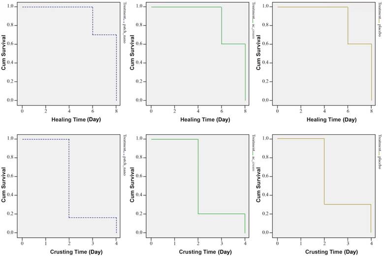
    A further study (Golestannejad, et al., 2021) was devised to investigate the effectiveness of the acyclovir-core nanofiber patches on living human subjects. Sixty participants who suffer from recurring herpes labialis were chosen for this trial. Participants were randomly split into three groups of twenty.  One group was treated with the acyclovir nanofiber patches, another group was treated with traditional topical acyclovir, and the third group was treated with a placebo nanofiber patch. Subjective pain symptoms were recorded using input from the subjects. Objective healing time and crusting time were recorded by the researchers using a pre-determined scale. The study resulted in a non-significant change in the healing and crusting time of the sores (Figure 4).

    Theoretically, the nanofiber patches should provide a more effective vector for the application of acyclovir cream. The effective surface area is vastly larger and should provide a vastly larger amount of medication to a more targeted area. The researchers in this study were not able to capitalize on the inherent benefits of the patches and would need to perform a follow-up study with a larger sample size, and an established metric on the amount of medication that will be administered via the use of these nanofiber patches. More research is required to find a middle ground between the initial study involving nanofiber patches, and the subsequent study (Golestannejad, et al., 2021).

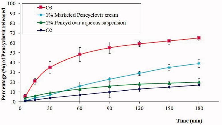
    Penciclovir is another of the most common antiviral drugs prescribed to help treat oral herpes. It can help reduce symptoms as well as shorten the healing time of cold sores. As effective as it is at treating oral herpes, it does not have good dermal permeability. Therefore, it needs to be formulated in relatively large dosages for creams and gels to overcome the low permeability. Current research into penciclovir involves the formulation of a penciclovir-loaded gel with better bioavailability and dermal permeability. Researchers have accomplished this by testing many different mixtures of an oil base, a surfactant, a self-nano emulsifying drug delivery system (SNEDDS), and the drug (Hosny, et al., 2021).

    Many essential oils were tested for the base, including lavender oil. Lavender oil is proven to have very strong antiviral activity and strong antiseptic properties. The ingredients for each gel were mixed using widely-available lab techniques, such as centrifuges, with a 6:4 ratio of oil and emulsion, along with the SNEDDS and 100mg of the drug. Each formulation was tested for droplet size and permeability. Permeability was tested by applying the gel to cultured buccal cells from sheep. The droplets needed to be large enough to maintain drug stability, and small enough to apply the drug to the cells. This was achieved most sufficiently by using the lavender oil formulation, which significantly outperformed every other formulation (Figure 5) (Hosny, et al., 2021).

    Relationship with Alzheimer’s disease:

                In 2021, Ruth Itzhaki introduced a review to the publication, Vaccines, in which she emphasizes her earlier work in 2017 which suggested an increased level of investigation into the relationship between HSV-1 and AD.  Since her 2017 publication, many researchers took up the call to scrutinize the two diseases in relation to one another (Itzhaki, 2021).  Many new and exciting research studies have been designed in the time since.  Such studies involve the use of stem cells to create human models of AD (Cairns, et al., 2020; D’Autio, et al., 2019; Abrahamson, et al., 2020).  Each of these studies have reinforced the suggestion that there is a strong relationship between HSV-1 and AD, however, none have yet pinpointed the exact mechanism by which these two diseases interact.

                The prevailing theory put out by Ruth Itzhaki, herself, is that HSV-1 can cause AD by creating an inflammatory response in the brain which creates more Aβ plaques and the accumulation of which creates a non-conductive environment in the neural cells of the brain.  Itzhaki surmises that the occurrence of a neural infection of HSV-1 later in life can increase the chances of an individual developing AD.  It has been observed that subjects who present with AD sometimes have HSV-1 in its latent form in their brains (Itzhaki, et al., 1997).  It was also observed that patients without AD have shown latent HSV-1 in their brains.  It is postulated by Itzhaki that the specific allele for apolipoprotein E4 (APOE4) is present in patients with AD who presented with a strong reactivation of latent HSV-1 in their brains later in life.  This allele is widely theorized to be the largest single factor in predicted a person’s risk for developing AD (NIH, 2023).  

    Itzhaki also notes that there is a wealth of clinical evidence showing that the treatment of a herpes-related virus prior to the onset of dementia or AD decreases the risk of developing symptoms.  For recent subjects who have received the shingles vaccine to prophylactically prevent a recurrence of the varicella zoster virus (VZV), there is a statistically significant reduction in risk of developing dementia symptoms later in life (Cairns, et al., 2022).  Additionally, one who is treated with daily valacyclovir (VCV) will also show a reduction in risk of developing dementia-related symptoms.

    This phenomenon could be due to Itzhaki’s theory that the virus, itself is causing AD but there could be another correlation that the researchers are not seeing.  For instance, there could be another neural virus that has not yet been detected, but is treatable with the same antivirals as HSV-1.  There could be many explanations for these correlations, but more research will need to be performed to find the necessary mechanisms behind the relationship.

                There are several genes associated with the development of AD, but the APOE4 gene seems to be the most prevalent of them.  The APOE4 gene is also associated with the development of cold sores.  There is no indication that having the APOE4 gene increases one’s risk for infection with HSV-1, however, subjects who do have the APOE4 gene and are infected with HSV-1 are more likely to present with cold sores (Itzhaki, 2022).  This observation seems to imply that the presence of the APOE4 gene increases the clinical manifestations of the virus, while, in others without the gene, clinical manifestations of the virus are less likely.  This explains why around 80% of most populations are infected with HSV-1, but only a small percentage of those subjects present with cold sores (Itzhaki, 2022).  It is estimated that somewhere between 15-25% of the population carries at least one allele for APOE4, but not all of those people develop AD, or HSV-1 (NIH, 2023).

    The scientific community is well-aware that HSV-1 can infect the brain, such is the case with herpes simplex encephalitis (HSE) (Patil, et al., 2022).  In order to test the correlation between HSV-1 and AD, a study was conducted in 2021 by Tran, et al.  Few pathogens can pass through the blood-brain barrier.  HSV-1 can pass through the blood-brain barrier and infect neural tissue.  The researchers in this study sought to determine the extent to which HSV-1 can contribute to symptoms of AD. When HSV-1 infects the brain, it can cause HSE.  

    The researchers theorized that frequent HSE flare-ups can create more Aβ plaques and NFTs in the brain, creating a typical neural environment that would be seen in a brain infected with AD.  The brain samples acquired were all from people who were diagnosed with AD and HSE.  The samples were stained and examined for HSV-1 infection.  Of the five samples, three showed Aβ plaques (Fig. 6). Two showed NFTs and all had HSE.  Through various imaging techniques, the researchers were able to observe the localization of HSV-1 cells around Aβ plaques.  Since only three of the five HSE samples had plaques present, it was determined that an active HSE infection is not associated with the exacerbation of AD symptoms.  

    This study was well-designed and was extremely successful at producing results.  The results were not what the researchers would have liked to have seen, however, they were extremely useful in the study of the association between HSV-1 and AD.  This is a weakness, as well as a strength of this research.  

    Not many original questions were answered by this study and there is no conclusive evidence that there is a relationship between HSV-1 and AD, however, this does open the door for future experiments regarding this relationship.  The sample size was quite small at five, but this was due to limited resources.  The researchers were able to extract as much information as was available from the few samples they were able to acquire.  Future studies should be designed to answer some of the open questions left after the completion of this study.  The researchers are still unsure of why there are HSV-1 particles localized around Aβ plaques when present.  This study was not able to answer the question of correlation between AD and HSV-1, but still created a strong basis for future endeavors into this profound research.

                Another study, published in 2020, sought to create a 3d human brain-like model and study the effects of HSV-1 on brain tissue as it relates to Alzheimer’s disease (AD).  This study was performed by Cairns, et al. and was based on a specific subset of AD cases.  Of the 6 million people in the United States who are infected with AD, 95% are diagnosed with late-onset AD.  Approximately 1-6% of cases are classified as early-onset AD (EOAD), in which a patient begins developing the disease under the age of 60 years old.  Patients diagnosed with EOAD exhibit the same hallmark symptoms of AD, including the development of β-amyloid plaques (Aβ) and neurofibrillary tangles (NFTs).  This form of AD is brought on by mutations in one of three specific genes associated with Aβ production.  

    Using EOAD as a model for the study is not ideal, as it is only present in 1-6% of AD cases.  A workaround developed for this study was to create a 3d brain-like model using human-induced neural stem cells (hiNSCs) which were cultivated using human foreskin fibroblasts.  The cultivated cells were then infected with HSV-1 which was titrated and purified to 2×10^7 PFU.  After infection, hiNSCs were shown to be highly infectable by HSV-1.  The infection of the human-like brain model resulted in increased gliosis and neural inflammation of hiNSCs. Regular treatment with valacyclovir (VCV) produced cell with significantly reduced gliosis and neural damage.  This study shows significant evidence that HSV-1 can cause some AD symptoms and can also create Aβ plaques characteristic of AD.  

    The researchers were extremely thorough in their notation and created an easily replicable study that has promising results.  As accurate an analog it is to human brain tissue, the 3d model is not a complete representation of a human subject.  Further research on human subjects is needed to determine the effects on living subjects of HSV-1.  The analog tissues were also not able to be fully accurate to an AD patient’s tissues.  A future study could use proposed aging methods on the tissues in order to more accurately model the brain of an AD patient.

    The results of this study suggest that there is a strong case for the treatment of AD by the use of antiviral drugs, such as VCV.  Recent trials of the use of the compound, GV971 have shown that the compound reduced neural inflammation and improved cognition in  trial subjects (Itzhaki, 2021).  Itzhaki believes that the use of a related, sulfated compound, fucoidan, could prove more successful in the treatment of AD.  This is due to the substances degree of sulfation, and the inherent antiviral properties of marine polysaccharides, from which the substance is derived.  These substances have been observed to reduce the incidence of Aβ plaques, which have been assumed one of the hallmark causes of symptoms of AD.  

    Originally published in 2006, a study conducted by Lesné, et al. concluded that the presence of Aβ contributed to cognitive decline in middle-aged mice.  The study uses mice of several age categories and measures their cognitive abilities, as well as their memory.  Researchers then examined the brains of the mice to find accumulations of Aβ plaques, and determined that the plaques were the cause of the decline in cognition with age.

    Several figures and images were used in the publication of this report, but one was thrust into question upon observations detected by Matthew Schrag (Pilier, 2022).  Schrag noticed that one of the images presented with apparent evidence of tampering.  This tampering implies that the research of Lesné, et al. may not have been entirely accurate.  It is also worth noting that Lesné’s experiment was not able to be replicated under peer-review by research groups outside of his own laboratory. 

    If Lesné‘s research was fabricated, then that could suggest that Aβ plaques are not as significant in the causation of symptoms of AD.  The apparent tampering of the photo alone does not suggest that the research is unsound.  However, if the research is unsound, then that could have contributed to the lack of advancements in the field of AD treatments over the last couple of decades, as most recent research has been designed to treat the symptoms and causes of Aβ plaques.

    The use of HSV-1 as an oncolytic virus:

    Herpes simplex virus type 1 is the only virus that is approved by the European EMA, and the American FDA as an oncolytic virus (OV). The viral genome is edited to utilize the virus as a directly injectable treatment for tumors. However, the strains used for synthesis are very specific strains normally used for laboratory reference. Talimogene laherparepvec (T-Vec) is the specific strain of HSV-1 that has been approved by the FDA for use as an OV (Koch, et al., 2020).  There are several other strains that are being developed and tested against numerous types of cancer cells.

    This marvel of modern medicine is achieved by genetically engineering a select strain of the virus to reduce its ability to infect neural cells and increase an immune response.  T-Vec, in particular, was engineered by researchers specifically to treat a form of melanoma.  To achieve this feat, researchers engineered the viral genome of a specific strain of HSV-1 so that it would present with lower neurovirulence, and augment an immune response.  They also inserted a gene that would further augment the immune response (Koch, et al., 2020).  The success of T-Vec has proven that HSV-1 can be genetically engineered to treat specific types of cancer.  In the wake of this success, many researchers across the world have designed studies and trials using other strain of HSV-1 to treat various types of cancer.

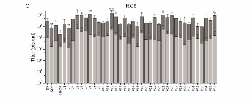
    A recent study performed by Kalke, et al. in 2022 tested the efficacy of thirty-six “wild” strains of HSV-1 for use as an OV.  Viral cells were lysed using a freeze-thaw cycle. Each of the thirty-six strains was tested for resistance to acyclovir before being tested on human cells.  Viral particles were used to infect Normal Human Corneal Epithelial cells (HCE) at 5 plaque-forming units (pfu) per HCE cell.  After genome sequencing, the viruses were each tested for oncolytic potential against several types of cancer cells at 2 pfu per cell.  The resulting infections successfully reduced the viability of cancer cells in neuroglioma, adenocarcinoma, and lymphoma (Fig. 8).  Especially effective was the infection of lymphoma cells, which saw a significant reduction in viability 2 days post-infection from six of the clinical strains of HSV-1 tested.  

    This study was designed to find new backbones for oncolytic HSV strains (oHSV).  The researchers were successful in reducing the viability of test cells representing various forms of cancer.  A weakness of this study is that it is somewhat unclear regarding numbers. In some sections, infections were said to have been incubated for two days, in some places four. Additionally, there does not seem to be any clinical explanation for the concentration of viral pfu’s used.  Another weakness of this study is that the cancer cells used were extremely limited.

    Kalke’s study was able to test many viral strains and prepare them for testing.  All strains were acquired regionally in Finland.  The researchers performed every test they needed in order to prove that their strains were viable as oncolytic viruses.  In the future, researchers will should seek to perform a full sequencing of the genomes of each strain, rather than just a few sections.  Ultimately, the results of this research can be instrumental in the design of future studies involving OVs.  With such a vast array or regional variants available, there are many possibilities of which strains to use for specific purposes.  

    Hematopoietic cells are characteristically resistant to HSV-1 because the infection mechanism of the virus does not typically target blood cells.  There have, however, been many cases of the sudden resolution of hematological malignancies following systemic viral infections documented.  It is theorized that this is because the systemic viral infection can also infect the cancer cells in a subjects bloodstream due to their differing microbiology from a normal hematological cell.

    If it is possible for a virus to kill these types of cancer cells, then it is possible to engineer an OV to treat them.  Aiming to investigate the efficacy of the usage of HSV-1 as an oncolytic vector for the treatment of such hematological malignancies, a study was designed by Ishino, et al. and published in 2020.  The researchers hypothesized that the key factor in the treatment of hematological malignancies was the expression of the nectin-1 receptor on the tumor cells. To test their theory, researchers used human cell lines derived from various hematological malignancies.

    The expression levels of the nectin-1 receptor were measured in each cell line. These tumor cells were implanted subdermally into mice on their left and right flanks and were then treated with a mock HSV-1 strain (T-01), and an ultraviolet-deactivated strain of T-01 (Fig. 9).  One mouse was treated via direct intratumor injection, while the other was treated without injection. The results showed a clear reduction in tumor growth in the mice who were treated with T-01.

    This proves that there is, in fact, a correlation between the expression of the nectin-1 receptor, and the oncolytic capabilities of T-01. This study showed some weaknesses in methods, but also some strengths. The researchers were able to test for many different cancer types.  However, the cancers tested were retrieved from patients who had recently relapsed, which could have shown a difference in the genome of the cancer cells.  A future trial would have to account for this by acquiring cancer cells from primary infections.  There were only a few mice tested and it was a very straightforward approach.  Another strength of this study is the extensive genome sequencing performed by the researchers.  This provides invaluable information for future research.   In the future, these data could be used to design a trial involving the treatment of hematological malignancies in human subjects.

    Conclusion:

             In my research, I have found that the treatments for HSV-1 have not had any real advancements in the decades since the development of acyclovir.  There have been several variants of this medication, but they are all most effective at treating the virus after a primary infection.  The ideal treatment for HSV-1 infections is a combination of pharmaceuticals and vitamins to prevent recurrent relapses, and ineffectual topical ointments after the prodromal phase has finished.  It is an inefficient way to treat the disease, not to mention expensive for the average consumer. 

                Without a better preventative treatment, or a cure, victims of the virus are at risk of developing other, more serious, presentations of the virus.  HSK is a very serious illness and one of the main causes of blindness in the United States.  In order to treat this disease and prevent the most debilitating symptoms, one is forced to take daily acyclovir for the remainder of their lives.  A daily acyclovir also just so happens to show a reduction in risk associated with the development of AD.

                Alzheimer’s disease is one of the most devastating diseases experiences by families in the United States today.  There has been little development in new treatments or cures for AD, as well.  More recent research has shown that there is a strong correlation between HSV-1 and AD under various circumstances.  Treatment of HSV-1 can reduce the risk of developing symptoms of AD.  HSV-1 viral particles have been observed in close proximity to Aβ plaques, which are one of the hallmark signs of AD.  There is a profound potential here to save millions of families the from the heartache of being forced to watch a loved one’s AD symptoms slowly increase in severity.  Further research into this relationship should be a high priority for labs across the world.

                Another disease that has no true cure and plagues families every day is cancer.  There are as many unique types of cancer as there are unique strains of HSV-1.  The most prevalent treatment today is a systemic poison that has horrible, long-lasting side effects.  With the development and use of HSV-1 oncolytic viruses, we could potentially treat many different types of cancer with a direct infection that could wipe out the tumor cells altogether.  Current trials involve the use of an oncolytic virus injected directly into tumors once every two weeks.  The OV is genetically engineered to infect the tumor cells, and not the neural cells of the host, reducing the risk of side effects to the patient.  This could prove to be a cheaper, safer alternative to the current chemotherapy treatments used in clinics around the world.

    What this research shows is that there is a profound need for more investigation into the HSV-1 virus for various reasons.  HSV-1 is a ubiquitous virus that shows up on all corners of the Earth and infects every rung of society.  Historically, it has not been thought to cause any significant long-term effects, but recent research shows that this is untrue.  Further research into the virus and its uses could prove invaluable to a society plagued with incurable diseases, such as cancers.  If we can learn to truly understand this ancient virus, we could unlock a cheat code to cure other diseases, as well as prevent the diseases caused by the virus.

    References:

    Abrahamson, E. E., Zheng, W., Muralidaran, V., Ikonomovic, M. D., Bloom, D. C., Nimgaonkar, V. L., & D’Aiuto, L. (2021). Modeling Aβ42 Accumulation in Response to Herpes Simplex Virus 1 Infection: 2D or 3D? Journal of Virology, 95(5)10.1128/JVI.02219-20

    Adams, Alex J., Klepser, Michael E. (2020). Pharmacy-Based Assessment and Management of Herpes Labialis (Cold Sores) with Antiviral Therapy . Innovations in Pharmacy, 11(3), 1-6. 10.24926/iip.v11i3.1532

    Alzheimer’s disease genetics: fact sheet (2004). . National Institute on Aging.

    Bodur, M., Toker, R. T., Özmen, A. H., & Okan, M. S. (2021). Facial colliculus syndrome due to a herpes simplex virus infection following herpes labialis. Turkish Journal of Pediatrics, 63(4), 727-730. 10.24953/turkjped.2021.04.023

    Cairns, D. M., Itzhaki, R. F., & Kaplan, D. L. (2022). Potential Involvement of Varicella Zoster Virus in Alzheimer’s Disease via Reactivation of Quiescent Herpes Simplex Virus Type 1. Journal of Alzheimer’s Disease, 88(3), 1189-12. 10.3233/JAD-220287

    Cairns, D. M., Rouleau, N., Parker, R. N., Walsh, K. G., Gehrke, L., & Kaplan, D. L. (2020). A 3D human brain–like tissue model of herpes-induced Alzheimer’s disease. Science Advances, 6(19), eaay8828. 10.1126/sciadv.aay8828

    Cunningham, A., Griffiths, P., Leone, P., Mindel, A., Patel, R., Stanberry, L., & Whitley, R. (2012). Current management and recommendations for access to antiviral therapy of herpes labialis. Journal of Clinical Virology, 53(1), 6-11. 10.1016/j.jcv.2011.08.003

    D’Aiuto, L., Bloom, D. C., Naciri, J. N., Smith, A., Edwards, T. G., McClain, L., Callio, J. A., Jessup, M., Wood, J., Chowdari, K., Demers, M., Abrahamson, E. E., Ikonomovic, M. D., Viggiano, L., De Zio, R., Watkins, S., Kinchington, P. R., & Nimgaonkar, V. L. (2019). Modeling Herpes Simplex Virus 1 Infections in Human Central Nervous System Neuronal Cells Using Two- and Three-Dimensional Cultures Derived from Induced Pluripotent Stem Cells. Journal of Virology, 93(9)10.1128/JVI.00111-19

    de Chadarevian, S., & Raffaetà, R. (2021). COVID-19: Rethinking the nature of viruses. History and Philosophy of the Life Sciences, 43(1), 2. 10.1007/s40656-020-00361-8

    Fiddian, A. P., Yeo, J. M., Stubbings, R., & Dean, D. (1983). Successful treatment of herpes labialis with topical acyclovir. Bmj, 286(6379), 1699-1701. 10.1136/bmj.286.6379.1699

    Golestannejad, Z., Khozeimeh, F., Mehrasa, M., Mirzaeei, S., & Sarfaraz, D. (2022). A novel drug delivery system using acyclovir nanofiber patch for topical treatment of recurrent herpes labialis: A randomized clinical trial. Clinical and Experimental Dental Research, 8(1), 184-190. 10.1002/cre2.512

    Hosny, K. M., Sindi, A. M., Alkhalidi, H. M., Kurakula, M., Alruwaili, N. K., Alhakamy, N. A., Abualsunun, W. A., Bakhaidar, R. B., Bahmdan, R. H., Rizg, W. Y., Ali, S. A., Abdulaal, W. H., Nassar, M. S., Alsuabeyl, M. S., Alghaith, A. F., & Alshehri, S. (2021). Oral gel loaded with penciclovir-lavender oil nanoemulsion to enhance bioavailability and alleviate pain associated with herpes labialis. Drug Delivery, 28(1), 1043-1054. 10.1080/10717544.2021.1931561

    Ishino, R., Kawase, Y., Kitawaki, T., Sugimoto, N., Oku, M., Uchida, S., Imataki, O., Matsuoka, A., Taoka, T., Kawakami, K., van Kuppevelt, T. H., Todo, T., Takaori-Kondo, A., & Kadowaki, N. (2021). Oncolytic Virus Therapy with HSV-1 for Hematological Malignancies. Molecular Therapy, 29(2), 762-774. 10.1016/j.ymthe.2020.09.041

    Itzhaki, R. F. (2021). Overwhelming Evidence for a Major Role for Herpes Simplex Virus Type 1 (HSV1) in Alzheimer’s Disease (AD); Underwhelming Evidence against. Vaccines (Basel), 9(6), 679. 10.3390/vaccines9060679

    Itzhaki, R. F., Lin, W., Shang, D., Wilcock, G. K., Faragher, B., & Jamieson, G. A. (1997). Herpes simplex virus type 1 in brain and risk of Alzheimer’s disease. The Lancet (British Edition), 349(9047), 241-244. 10.1016/S0140-6736(96)10149-5

    Iwanaga, J., Fukuoka, H., Fukuoka, N., Yutori, H., Ibaragi, S., & Tubbs, R. S. (2022). A narrative review and clinical anatomy of herpes zoster infection following COVID‐19 vaccination. Clinical Anatomy (New York, N.Y.), 35(1), 45-51. 10.1002/ca.23790

    Kalke, K., Orpana, J., Lasanen, T., Esparta, O., Lund, L. M., Frejborg, F., Vuorinen, T., Paavilainen, H., & Hukkanen, V. (2022). The In Vitro Replication, Spread, and Oncolytic Potential of Finnish Circulating Strains of Herpes Simplex Virus Type 1. Viruses, 14(6), 1290. 10.3390/v14061290

    Kazsoki, A., Palcsó, B., Alpár, A., Snoeck, R., Andrei, G., & Zelkó, R. (2022). Formulation of acyclovir (core)-dexpanthenol (sheath) nanofibrous patches for the treatment of herpes labialis. International Journal of Pharmaceutics, 611, 121354. 10.1016/j.ijpharm.2021.121354

    Kim, H., Kang, K. W., Kim, J., & Park, M. (2022). Uncommon cause of trigeminal neuritis and central nervous system involvement by herpes labialis: a case report. BMC Neurology, 22(1), 1-294. 10.1186/s12883-022-02823-x

    Koch, M. S., Lawler, S. E., & Chiocca, E. A. (2020). HSV-1 Oncolytic Viruses from Bench to Bedside: An Overview of Current Clinical Trials. Cancers, 12(12), 3514. 10.3390/cancers12123514

    Öztekin, A., & Öztekin, C. (2019). Vitamin D Levels in Patients with Recurrent Herpes Labialis. Viral Immunology, 32(6), 258-262. 10.1089/vim.2019.0013

    Patil, A., Goldust, M., & Wollina, U. (2022). Herpes zoster: A Review of Clinical Manifestations and Management. Viruses, 14(2), 192. 10.3390/v14020192

    PEDRAZINI, M. C., ARAÚJO, V. C., & MONTALLI, V. A. M. (2018). The effect of L-Lysine in recurrent herpes labialis: pilot study with a 8-year follow up. RGO – Revista Gaúcha De Odontologia, 66(3), 245-249. 10.1590/1981-863720180003000083517

    Pilier, C. (2022). BLOTS ON A FIELD? A neuroscience image sleuth finds signs of fabrication in scores of Alzheimer’s articles, threatening a reigning theory of the disease. Science (American Association for the Advancement of Science), 377(6604), 358. https://www.science.org/content/article/potential-fabrication-research-images-threatens-key-theory-alzheimers-disease

    Ramalho, K. M., Cunha, S. R., Gonçalves, F., Escudeiro, G. S., Steiner-Oliveira, C., Horliana, A. C. R. T., & Eduardo, C. d. P. (2021). Photodynamic therapy and Acyclovir in the treatment of recurrent herpes labialis: A controlled randomized clinical trial. Photodiagnosis and Photodynamic Therapy, 33, 102093. 10.1016/j.pdpdt.2020.102093

    Rujescu, D., Herrling, M., Hartmann, A. M., Maul, S., Giegling, I., Konte, B., & Strupp, M. (2020). High-risk Allele for Herpes Labialis Severity at the IFNL3/4 Locus is Associated With Vestibular Neuritis. Frontiers in Neurology, 11, 570638. 10.3389/fneur.2020.570638

    Silva-Alvarez, A. F., de Carvalho, A. C. W., Benassi-Zanqueta, É, Oliveira, T. Z., Fonseca, D. P., Ferreira, M. P., Vicentini, F. T. M. C., Ueda-Nakamura, T., Pedrazzi, V., & de Freitas, O. (2021). Herpes Labialis: A New Possibility for Topical Treatment with Well-Elucidated Drugs. Journal of Pharmaceutical Sciences, 110(10), 3450-3456. 10.1016/j.xphs.2021.06.029

    Tran, D. N., Bakx, A. T. C. M., van Dis, V., Aronica, E., Verdijk, R. M., & Ouwendijk, W. J. D. (2021). No evidence of aberrant amyloid β and phosphorylated tau expression in herpes simplex virus-infected neurons of the trigeminal ganglia and brain. Brain Pathology (Zurich, Switzerland), 32(4), e13044-n/a. 10.1111/bpa.13044

    Treister, N. S., & Woo, S. (2010). Topical n-docosanol for management of recurrent herpes labialis. Expert Opinion on Pharmacotherapy, 11(5), 853-860. 10.1517/14656561003691847

    Wang, L., Wang, R., Xu, C., & Zhou, H. (2020). Pathogenesis of Herpes Stromal Keratitis: Immune Inflammatory Response Mediated by Inflammatory Regulators. Frontiers in Immunology, 11, 766. 10.3389/fimmu.2020.00766

    Zhu, S., & Viejo-Borbolla, A. (2021). Pathogenesis and virulence of herpes simplex virus. Virulence, 12(1), 2670-2702. 10.1080/21505594.2021.1982373

    Appendix:Diagram

Description automatically generated

    Figure 1: Herpes virion containing a large, double-stranded DNA genome contained and protected by a capsid, which is surrounded by a tegument layer.  The envelope contains glycoproteins which are used to attach to host cells and release genetic material. (Zhu and Viejo-Borbolla, 2021). 

    Figure 2: HSV cell cycle. 1: Viral glycoproteins bind to receptors on the cell plasma membrane. 2: The viral capsid fuses with the plasma membrane of the cell. 3: Viral capsid and proteins from the integument are transported to the cell nucleus via microtubules. 4, 6, 9: Transcription of the viral genome takes place in the cell nucleus. 5, 7, 10: Translation of the viral genome. 8: Viral DNA replication. 11: Replicated viral DNA undergoes encapsidation. 12: Capsid is transported through the cytoplasm. 13: Vesicles transport the capsid to the plasma membrane 14: Viral capsid exits the cell (Zhu and Viejo-Borbolla,2021).

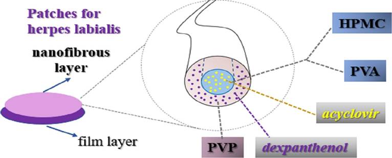
    Figure 3: Acyclovir core, PVP sheath nanofiber used to create medicated patches. Acyclovir hydrochloride and dexpanthenol were used as active pharmaceutical ingredients. Polyvinyl alcohol (PVA) and hydroxypropyl methylcellulose (HPMC) were used as the precursor solutions for formulation. Polyvinylpyrrolidone (PVP) was used to formulate the shell (Kazsoki, et al., 2022).

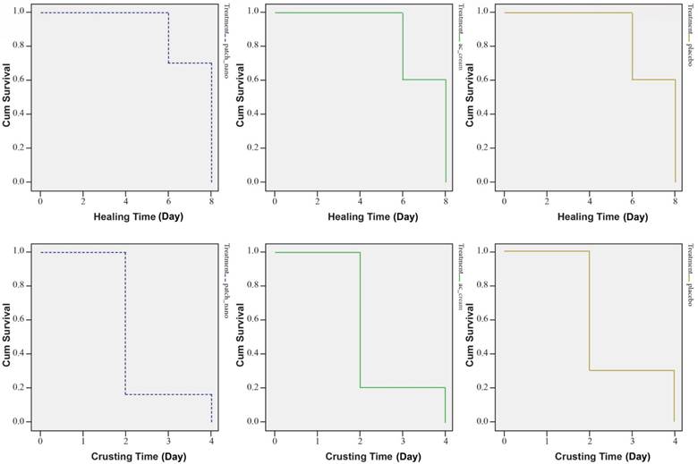
    Figure 4: Survival curves of crusting and healing time in subjects’ lesions. Groups from left to right are acyclovir nanofiber patches, topical acyclovir, and placebo. The mean crusting time for each group, respectively was 2.3, 2.4, and 2.6 days. the mean healing time was 7.4, 7.7, and 7.2 days, respectively (Golestannejad, et al., 2021).

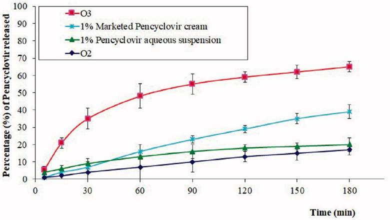
    Figure 5: Percentage of acyclovir released per minute with differing application vectors. O2: chitosan hydrogel loaded with PV powder, O3: chitosan hydrogel loaded with optimized PV-LO SNEDDs , 1%: PV cream, and 1% PV aqueous suspension (Hosny, et al., 2021).

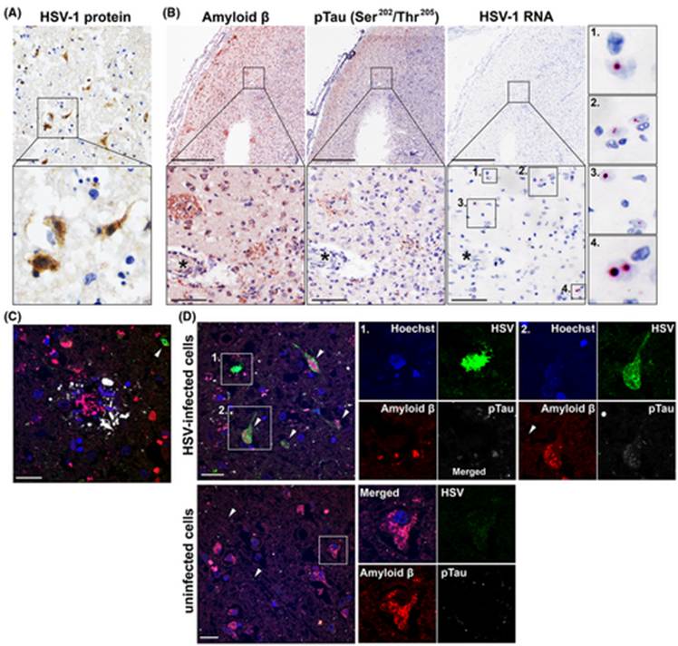
    Figure 6: Various imaging techniques showing the proximity of HSV infected cells to Aβ plaques. Proteins are stained with immunohistochemistry (IHC) A: Brain sample stained for HSV proteins. B: Consecutive samples showing staining for pTau and Aβ proteins. C, D: samples stained with immunofluorescence for HSV, pTau, Aβ. Cell nuclei are stained in blue, HSV proteins are stained green, Aβ is red, and pTau is white. These images prove the spatial correlation between HSV1 and AD signs (Tran, et al., 2021).

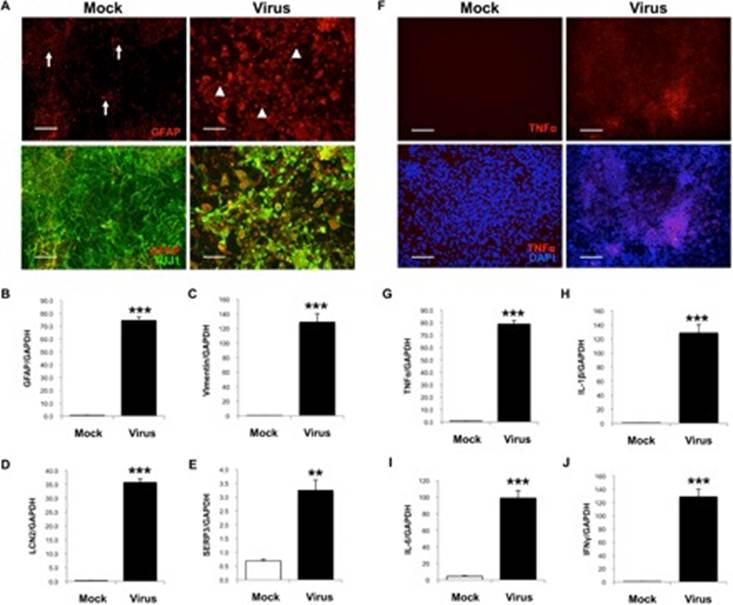
    Figure 7: Immunostaining images showing that HSV-1 infected mock-brain cells exhibit reactive gliosis and inflammation.  A, B: hiNSCs infected with HSV-1 showing signs of neuroglial activity, indicative of gliosis. C-J: Several other inflammatory markers present in cases of AD are found in hiNSCs infected with HSV-1 (Cairns, et al., 2020).

    Figure 8: Graph showing the intracellular and extracellular titers of each clinical isolate. HCE cells were infected at 0.1 pfu/cell and titers were measured at pfu/ml. Each of the 36 clinical isolates were tested and the light grey bars show the extracellular viral titer measured.  The dark grey bar shows the intracellular titer, which is consistently much higher than the extracellular titers.  This shows the high intracellular virulence of the virus.

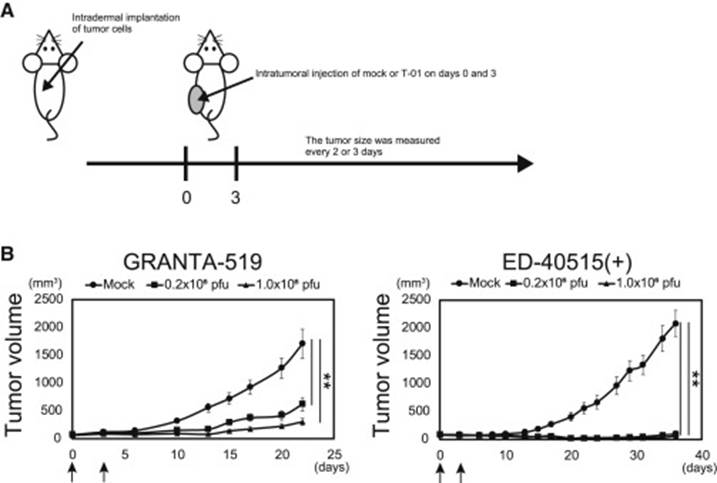
    Figure 9: Anti-tumor effects of test mice.  A: The treatment plan for the mice. B: growth curve of tumors after treatment. The arrows mark the dates of the injections on day 0 and day 3. The size of each tumor was measured every 2-3 days.

  • SPAGHETTI was a program designed to generate simulations of genetic populations with specific segregating molecular markers, incorporating realistic complications such as duplicated loci and segregation distortion (Tinker, 2010). It generated output compatible with standard genetic mapping software and facilitated the testing and demonstrating linkage map construction methods. The software, source code, sample files, and instructions were freely available for public use.

    The need for SPAGHETTI arose from the increasing ease of generating genetic linkage maps, which could mask inaccuracies. While modern genomics relied on high-quality maps, even experienced researchers could produce flawed maps. SPAGHETTI addressed this by allowing users to simulate genetic data with known parameters, providing a way to evaluate mapping methodologies objectively.

    SPAGHETTI operated via a command line or simple graphical interface, requiring one or two input files. The primary input file specified population parameters (e.g., population type, size, the proportion of segregating loci, missing/mis-scored data, and markers with reversed scoring phase). It supported simulations of F2, doubled haploid, and recombinant inbred populations. A second input file defined the underlying genome, specifying locus names, positions, and other optional parameters. If a user does not provide a genome file, SPAGHETTI could generate a random genome, which could be saved and modified for future use.

    The program produced two output files for each simulated population: a “perfect” dataset with all loci scored codominant and without errors and a dataset incorporating the specified abnormalities. These files were output in formats compatible with Mapmaker and JoinMap software. A log file recorded data abnormalities, offering transparency. The simulation also could add random error or “noise” to the process (Tinker & Mather, 1993).

    Figure 1: SPAGHETTI simulation (Tinker, 2010).

    In this example of use, SPAGHETTI simulated five F5 recombinant inbred populations with a 100 cM chromosome. JoinMap analysis shows that the single population map (left) had fewer markers than the merged map (right). Researchers evaluated the accuracy by comparing the marker positions to the original genome with a mean squared error difference. These results show the benefits of consensus mapping and demonstrate that the original population sizes were too small for aping (Figure 1) (Tinker, 2010).

    References

    Tinker, N. A. (2010). SPAGHETTI: Simulation software to test genetic mapping programs. Journal of Heredity, 101(2), 257-258. https://doi.org/10.1093/jhered/esp114

    Tinker, Nick & Mather, Diane. (1993). GREGOR: Software for Genetic Simulation. Journal of Heredity. 84. 237. 10.1093/oxfordjournals.jhered.a111329.

  • Rett syndrome (RTT) is a severe neurodevelopmental disorder primarily affecting women, caused mainly by mutations located on the MECP2 gene found on the X chromosome (Merritt, 2020). MECP2 is a gene that encodes the methyl-CpG binding protein 2 (MeCP2), a regulator of gene expression in the brain. MeCP2 binds to methylated DNA sequences, impacting the activity of numerous genes that are sed in neuronal development and function (Chahrour & Zoghbi, 2007). Understanding the types of mutations in MECP2 that cause RTT, compared to those found in the general population, can provide insight into how genetic variations can lead to different outcomes.

    Genetic testing confirming an MECP2 mutation helps diagnose Rett syndrome. The mutations responsible for RTT are typically those that severely disrupt the function or production of the MeCP2 protein (Kyle, Vashi, & Justice, 2018). The most common mutations identified in individuals with RTT are missense mutations with an expected single nucleotide variant count of 229.3 vs. the observed count of 281. The mutations causing Rett syndrome result in a substantial loss or impairment of MeCP2 protein function, disrupting its role in brain development and maintenance (Kyle, Vashi, & Justice, 2018).

    To understand the baseline variation in the MECP2 gene, large genomic databases like the Genome Aggregation Database (gnomAD) are examined. Analyzing the non-neuro subset of gnomAD shows a different spectrum of MECP2 mutations than RTT patients. While missense, synonymous, and intronic variants are observed, the frequency of highly disruptive nonsense and frameshift mutations is much lower in this population. Variants found here generally occur at very low allele frequencies. The overall pattern suggests that MECP2 variants common in the general, non-neuro population are less likely to be severely damaging. There is a higher relative prevalence of synonymous variants. Missense variants are predicted to be benign or likely benign. However, this doesn’t mean pathogenic variants are absent; some, like p.Arg115His, are found at very low frequencies, possibly indicating incomplete penetrance or variable expressivity. Allele frequency aids pathogenicity assessment; the relatively high frequency of p.R344W in South Asians led to its classification as likely benign. While the gnomAD non-neuro population harbors various MECP2 variants, the spectrum is enriched for benign or likely benign changes compared to the pathogenic mutations causing RTT, though rare potentially pathogenic variants exist.

    Using the gnomAD 2.1.1 database, I was able to locate four pathogenic or likely pathogenic mutations for Rett syndrome. The first pathogenic variation (Variation ID: 143384) is a frameshift mutation, with several submissions on ClinVar alluding to its pathogenicity. The second is a missense mutation (Variation ID: 2505626). The third located mutation is another frameshift mutation (Variation ID: 143360). The final mutation (Variation ID: 11844) had the most highly rated classification of the four, although it is classified as “likely pathogenic” rather than “pathogenic.”

    The different mutations within the MECP2 gene lead to diverse outcomes resulting from the relationship between a gene, its protein product’s structure, and function. A protein’s function depends on its shape, which an amino acid sequence determines (Chahrour & Zoghbi, 2007). If there is a mutation in the amino acid sequence, the protein will be disfunctional. The location of a mutation is important, meaning that changes within functional domains are likely to disrupt the protein’s function, which can cause disease (Chahrour & Zoghbi, 2007). Mutations in less important regions may have little impact. Nonsense and frameshift mutations often cause a loss of function and disease. Missense mutations have variable effects depending on the specific amino acid change and location. Synonymous mutations are usually benign but can occasionally affect mRNA processing.

    Other factors, like genetic background or, in women, the pattern of X-chromosome inactivation, can influence the outcome (Chahrour & Zoghbi, 2007). A mutation’s likelihood of causing disease depends on how and where it affects the protein. Changes that compromise essential structure or function are classified as pathogenic. Those with less functional impact are classified as benign or likely benign.

    Variation IDType of MutationClassification
    11844missense_variantLikely pathogenic
    2505626frameshift_variantPathogenic
    2505626missense_variantPathogenic
    143360frameshift_variantPathogenic/Likely pathogenic

    Table 1: Variation ID, mutation type, and classification of all pathogenic or likely pathogenic mutations for Rett syndrome

    Figure 1: gnomAD analysis of MECP2 genetic mutations.

    Figure 2: ClinVar and gnomAD variants of MECP2 gene, filtered for pathogenic/likely pathogenic mutations.

    References

    Chahrour, M., & Zoghbi, H. (2007). The Story of Rett Syndrome: From Clinic to Neurobiology. Neuron, 56(3), 422–437. https://doi.org/10.1016/j.neuron.2007.10.001.

    Kyle, S., Vashi, N., & Justice, M. (2018). Rett syndrome: a neurological disorder with metabolic components. Stephanie M. Kyle, Neeti Vashi and Monica J. Justice, 170216. https://doi.org/10.1098/rsob.170216.

    Merritt, J. (2020). On the mechanisms governing Rett syndrome severity. UC San Diego Electronic Theses and Dissertations, https://escholarship.org/uc/item/41t7g3m6.

  • Landscape genomics is an interdisciplinary field that merges population genetics, landscape ecology, and spatial statistics. The goal of landscape genomics is to understand how environmental factors affect the genetic makeup of populations. The field seeks to uncover the genetic basis of adaptation by linking specific genetic variants to ecological pressures. This can provide powerful insights into evolutionary processes in a real-world context.

    Landscape genomics is broadly divided into two main conceptual frameworks, neutral and adaptive. Neutral landscape genomics, also known as landscape genetics, focuses on how the landscape influences demographic processes. It utilizes neutral genetic markers, which are not subject to natural selection. This approach examines how factors such as geographic distance or physical barriers limit gene flow. It also helps to understand population structure and history.

    The goal of adaptive landscape genomics is to detect the signature of natural selection. This approach utilizes genome-wide data to identify specific genes that are strongly associated with environmental variables. By identifying these outlier loci, researchers can pinpoint the genetic basis of local adaptation. This process allows them to test for isolation by environment.

    Two main types of analytical methods are used to achieve these goals. Distance-based analyses are often used in neutral studies. These methods work by correlating different matrices of data. For example, a matrix of genetic distances is compared to a matrix of geographic distances. This type of analysis determines whether genetic patterns are better explained by spatial separation or environmental differences. Correlative approaches are known as genotype-environment association methods. These are the primary tools of adaptive landscape genomics. The statistical models directly test for associations between individual genetic loci and particular environmental variables. A strong correlation suggests the locus may be under selection.

    Landscape genomics has numerous practical applications. It is used to predict a species’ vulnerability to climate change. It helps to identify resilient populations for conservation and guide genetic rescue efforts. This field can also inform the selection of climate-adapted crops for agriculture. Detecting polygenic adaptation is statistically difficult because many genes of small effect control these traits. It is also hard to distinguish the signals of selection from complex demographic history. Moving from a statistical association to experimentally validating the function of a candidate gene is a major hurdle. Validation requires a substantial amount of time and resources.

    References

    Rajora, O. P. (2019). Population Genomics: Concepts, Approaches and Applications 1st ed. Springer Nature Switzerland AG.

  • Nicholas Holmes#, Patrick Muller*, Rishi Patel%, Anisha Tehim&, Atharva Imamdar&, Saachi Yadav&, Sharon Alex&, Vibha Narasayya& and Vinayak Mathur#

    # Department of Science, Cabrini University, Radnor, PA 19087
    *Eurofins, Chester Springs, PA 19425

    %University of Pennsylvania, Philadelphia, PA 19104

    &Penn Summer Prep Program, Philadelphia, PA 19104

    Abstract

    Horizontal gene transfer (HGT) plays a beneficial role in the evolution and survival of bacteriophages and bacteria. The extent of HGT between Streptococcus bacteria and associated bacteriophages, focusing on viral major capsid proteins, was studied utilizing a bioinformatics approach. Evidence of HGT was identified via the community science analysis pipeline and the BLAST database. Evolutionary relationships were assessed using MEGA software to construct phylogenetic trees. Overall relationships were then represented as networks via the Gephi application. Literature has shown that the major capsid protein in bacteria works analogously to bacterial microcompartments, protecting genetic materials and organelles. These observations, as well as genomic locations of genes coding for major capsid proteins, DNA polymerases, DNA topoisomerases, and other associated molecules, have led to their uses as biomarkers of potential HGT cases. The results provide evidence of extensive HGT between bacteria and bacteriophages, which helps in understanding their evolution and potential therapeutic uses.

    Introduction

    Antibiotic resistance within bacterial populations is rising to dangerously high levels and new resistance mechanisms are emerging and spreading globally. One such mechanism that has been observed is horizontal gene transfer (HGT). HGT occurs when genetic material is exchanged between organisms in a non-genealogical manner (Goldenfeld and Woese, 2007). This genetic exchange is unlike the genetic exchange which occurs from parent to offspring, as HGT usually occurs between different organisms which are not related. Through the means of HGT, a bacteria can pick up many different functions, including antibiotic resistance and virulence factors. (Deng et al. 2019)

    Bacteriophages have shown to have a role in transfer of genetic material between bacteria (Borodovich et al., 2018). Bacteriophages can infect bacteria either through the lytic cycle (called lytic phages) or the lysogenic cycle (called temperate phages) (Rehman et al., 2019). The lysogenic cycle involves the bacteriophage integrating their genome into that of the host cell, and can become dormant, only to infect the cell when it undergoes activation (Labonté et al., 2019). These temperate phages can serve as vectors for HGT between bacterial species via transduction (Labonté et al., 2019). Phage transduction can be studied by examining a bacterial genome and locating the pockets of viral DNA. Phages have effects on the control of bacterial populations, the spread of virulence factors and antibiotic resistance genes, resulting from unique combinations of genetic diversity (Cumby et al., 2015). The mechanism by which these viruses infect bacteria and how these drive their evolution is poorly understood and is crucial to understand where they originated from (Cumby et al., 2015). Bacteriophages have unique host range, and their specificity is determined by their specific structures to attach to specified host bacterial cell receptors and infect the cells.

    The protein that we focus on in our study is the major capsid protein. Capsids are the morphological structures that contain the condensed form of the genetic material of a bacteriophage, and also protect it from any outside physical and chemical damages. Recent research has shown that mutations in the genes that code for these proteins are necessary for certain interactions with different host cell receptors and appear to contribute to the stability of a given capsid. This suggests that these mutations aid in broadening bacteriophages’ host ranges (Labrie et al., 2014).

    Interestingly, there are similarities between encapsulin proteins that form bacterial structures resembling shells, and the major capsid protein of the HK-97 bacteriophage (Freire et al., 2015). Structural identities are also seen with capsid proteins and S-layer lattice protein components of the cell envelope of prokaryotes and bacteria (Freire et al., 2015). Fusogenic proteins of enveloped viruses which enable the fusion between them and host cell membranes, have also been shown to function analogously to the SNARE family of proteins of Caenorhabditis elegans, which encode for fusion of intracellular vesicles to their cell membranes and allow for cell–to–cell communication (Freire et al., 2015). Not only that, the structures and functions of major capsid proteins and bacterial microcompartments are very similar. Bacterial microcompartments are protein shells that encase enzymes, molecules essential for the microorganism, and may be even their genetic material, protecting these items from degradation or physical or chemical damages in or out of the cell (Krupovic & Koonin, 2017). These notions provide insight into how the ideas and concepts of these proteins encoding capsids that protect their genetic material have expanded from beyond unique to bacteriophages, but to other microorganisms as well.

    Bioinformatics analyses and studies have demonstrated bacteria that have undergone HGT with bacteriophages possess conserved genomic regions pertaining to not just to single genes, but multiple, different genes that are located relatively close to one another. According to research presented by Sabath et al., 2012, overlapping genes and sequences are very common in viral genomes. Expressions of these genes have been confirmed, while functionality requires further investigation. Interestingly, the resulting, translated proteins lack a stable, tertiary, three-dimensional structure characteristic of most normal, wild-type proteins (Sabath et al., 2012). In addition to genes encoding major capsid proteins, genes that encode for DNA polymerases, DNA topoisomerases, and other molecules associated with genome replication are inherited through HGT as well. These genes have been shown to be located close together within viral genomes. Appropriately, these genes are termed genomic islands, which refer to groups of unique open reading frames that contain sequences that encode given traits or carry out specific functions (Villa & Viñas, 2019). Examples of genomic islands that have been investigated carry out functions pertaining to virulence and pathogenicity, symbiosis, metabolism, fitness, and antibiotic resistance (Finke et al., 2017). The specific mechanism of HGT involved in how these genomic islands are integrated into the genomes of host organisms remains unknown (Villa & Viñas, 2019). This property provides an opportunity to utilize major capsid proteins as biomarkers when analyzing genomic sequences to establish evidence of HGT amongst bacteriophages and bacteria (Born et al., 2019).

    In this study we focused on the major capsid protein in Streptococcus genus of bacteria. Studying how bacteria acquires its resistance to antibiotics is necessary, due to the shrinking list of effective antibiotics. The objective of this study is to assess the extent of HGT between bacteria species and associated bacteriophages, using the major capsid proteins as biomarkers. Our results indicate that overlapping open reading frames composed of varying numbers of base pairs are located close to the major capsid protein in the bacterial genomes. Whether or not these regions encode functional proteins is not entirely known. Current annotations present in databases indicate that these genomic regions encode several different proteins that contribute to the structure and morphology of the major capsid protein (Rosenwald et al., 2014).

    Methods

    HGT & Community Science Project Pipeline

    Positive cases of HGT between bacteriophages and bacteria were determined using the Community Science Project Pipeline (Mathur et al., 2019). A list of accession numbers of bacteriophage major capsid proteins was generated from the NCBI database. Each phage accession number was searched against the bacteria database on NCBI using BLASTp to generate positive hits (referred to as Forward BLAST) (Johnson et al., 2008). The top 10 hits based on the cut-off criteria of e-values of 1e-50 or lower, and a query coverage of 70% or higher, were recorded. The top bacterial hit accession number was then searched against the Virus database on NCBI using BLASTp (referred to as Reverse BLAST) (Johnson et al., 2008). Again, the top 10 hits which satisfied the cut-off parameters were recorded. If the top virus hit accession number in the Reverse BLAST matched the original virus accession number query, that bacteria-virus pair was recorded as a potential positive case of HGT. In total, 75 phage accession numbers were tested to give 21 positive HGT bacteria-virus pairs (Table 1).

    Evolutionary History of Bacteria and Bacteriophages – Comparative Genomics

    The evolutionary history of bacteria and bacteriophages was assessed via comparative genomics. FASTA sequences of the major capsid protein from all positive cases of HGT were uploaded to MUSCLE software and aligned (Edgar, 2004). (Supplementary Figure 1). These sequences were then uploaded to the MEGA7 software to generate phylogenetic trees (Kumar et al., 2015). The phylogenetic tree was constructed based on maximum likelihood method and bootstrapping value of 100, seen in Figure 1. Based on the results, the Streptococcus clade of bacteria was selected for further analyses.

    Synteny & Evolutionary Relationships

    The Streptococcus clade of bacteria and bacteriophages were selected for the synteny analysis. Synteny for the Streptococcus clade of bacteria and bacteriophages was determined using the software MAUVE (Darling et al., 2010). Major capsid gene sequences were downloaded from NCBI for both bacteria and bacteriophages. The Mauve synteny output was generated for all the phages, bacteria and the visualization of the bacteria and phage sequence.

    Gephi Network Analysis

    The top six results from the Forward and Reverse BLAST searches were collected based on the Community Science Pipeline and were each organized as a node into the Gephi software for network analysis (Bastian et al., 2009). Connections between bacteria and bacteriophages based on the generated phylogenetic trees were input into Gephi as edges. A node in the center of the network with the most edges connected to it was indicative of the ancestral sequence that was shared by most bacteria and bacteriophages through HGT.

    Results

    Comparative Genomics

    Based on the arrangement on the phylogenetic tree and validation by the bootstrapping values, there is a high likelihood that Streptococcus bacteriophages and bacteria were involved in HGT with respect to the major capsid protein. Despite the major capsid protein being present in the five bacteriophages and bacteria, their location in the genomes of each species varies. This suggests mutations such as translocations and insertions have occurred over time (Kyrillos, et al., 2016). This could explain the divergence of pairs of bacteriophages and bacteria in the phylogenetic tree. One pair that is the most divergent and in its own unique clade and not associated with the other pairs is the connection between the Streptococcus phage VS-2018a and the major capsid protein E in Streptococcus thermophilus.This is also reflected in their MUSCLE alignments that vary compared to the other bacteria and bacteriophage pairs.

    Synteny & Evolutionary Relationships

    The Mauve software was used to create a multiple sequence alignment and predict synteny of Javan Streptococcus bacteriophage and bacteria pairs using the progressive Mauve algorithm. In the synteny map of the four Javan prefixed bacteriophages, the major capsid protein lies in the range of approximately 500-2000 base pairs. (Supplementary Figure 3). There is a consistent alignment based on the peak height and coloration patterns with Phage VS2018 having the most unique genome arrangement. The S.thermophilus is missing a 400 base pair region upstream of the major capsid protein gene, as indicated by a shift in the sequence alignment (Supplementary Figure 4). The five phage sequences of interest are in reverse orientation in the genome indicated by the peaks falling below the main sequence line in Figure 2. The area between 850-970 base pairs is a unique region found only in S.thermophilus bacteria and the phage VS 2018a pair. This is expected as this pair lies on a separate clade in the phylogenetic tree generated previously. The alignment of the genomes indicates that the region upstream and downstream of the major capsid gene is also shared between these bacteria and bacteriophage pairs. This pattern indicates that there is not just the major capsid gene that is shared between bacteria and phages but instead a whole chunk of the genome.

    Gephi Network Analysis

    The central node in the network corresponds to a hypothetical protein in Streptoccoccus pyogenes. As seen in the top six results of the Reverse BLAST, this node has multiple shared edges with a major capsid protein in Streptococcus bacteriophages Javan 146, 454, 464, 474, 459, 484, 166 (Figure 3). The generated network shows that the connections are the same as they appear in the phylogenetic tree. The central node of the gene encoding a hypothetical protein in S.pyogenes connects closely to different strains of itself and a Javan bacteriophage 464. This relationship suggests that those two bacteria and bacteriophage pairs could be where the initial transfer of genomic material had occurred.

    Discussion

    HGT of the major capsid protein has allowed for Streptococcus bacteriophages and bacteria to display survival of the fittest to survive in constantly changing environments. In doing so, greater genetic diversity is achieved through HGT, thus potentially speeding up adaptation and overall evolution.

    Upon review of the scientific literature, the role and functions of major capsid proteins

    could potentially serve as a bacterial microcompartment protecting the bacterial genome from

    physical and chemical damages akin to the functions of viral capsids, as a result of HGT (Krupovic & Koonin, 2017). Interestingly, there are similarities in morphology

    between the S-layer lattice proteins present in mostly archaea bacteria. Based on this notion,

    perhaps these proteins function as a means to protect the genetic material of archaea from

    physical or chemical damages. Perhaps even archaea evolved to possess such as a structure from

    HGT as a means to survive in its native environment of hot springs or areas of high temperature

    (Freire et al., 2015).   

    It has been suggested that bacterial genes acquired through HGT are usually quickly deleted from the genome unless they are to be utilized for some specific reasons later on (Rosenwald et al., 2014). For example, genes acquired through HGT that improve metabolism in bacteria can be expressed under given circumstances. It is upon changes in the environment or medium that render these genes functionless that can result in the deletion of the genes. This is understandable as bacterial genomes tend to be very compact and constituent (Moran, 2002). This most likely occurs as a means to conserve internal energy by not expressing genes that are not necessary or will not be considered as such. These notions can be both determined via RNA sequencing techniques in which the protein of interest is isolated, its messenger RNA is extracted and purified from other RNA molecules, for study (Rosenwald et al., 2014).

    By identifying HGT within the major capsid protein sequences for the Streptococcus bacteria and the bacteriophages that infect them, we can begin to understand the extent of HGT within bacterial populations. We propose that the major capsid protein can be used as a biomarker to identify HGT in other bacteria species as well. There is evidence to suggest that when genes are transferred horizontally, it is not just a single gene but a whole genomic region consisting of multiple genes (Szöllősi et al., 2015). A future direction of this research would be to identify the gene regions flanking the major capsid protein in the bacterial genome and understand the functionality of those genes and the role they play within the bacteria.

    In this study, we focused on the Streptococcus genus, but it can easily be expanded to include a larger dataset of bacteria and bacteriophage pairs based on data availability in the NCBI database. It is imperative to study the extent and rate of HGT in bacterial populations as it is a key mechanism for bacteria to acquire antibiotic resistance genes, and thus has implications for human health worldwide.

    References

    Bastian, M., Heymann, S., & Jacomy, M. (2009). Gephi: an open-source software for exploring and manipulating networks. Print. N. P. Retrieved from https://gephi.org/users/publications/

    Born, Y., Knecht, L. E., Eigenmann, M., Bolliger, M., Klumpp, J., & Fieseler, L. (2019). A major-capsid-protein-based multiplex PCR assay for rapid identification of selected virulent bacteriophage types. Archives of Virology, 164(3), 819–830. doi: 10.1007/s00705-019-04148-6.

    Borodovich, T., Shkoporov, A. N., Ross, R. P., & Hill, C. (2018). Phage-mediated horizontal gene transfer and its implications for the human gut microbiome. Research in Microbiology, 169(7-8), 366-373. https://doi.org/10.1016/j.resmic.2018.04.005

    Cumby, N., Reimer, K., Mengin‐Lecreulx, D., Davidson, A. R., & Maxwell, K. L. (2015). The phage tail tape measure protein, an inner membrane protein and a periplasmic chaperone play connected roles in the genome injection process of E. coli phage HK97. Molecular Microbiology, 96(3), 437-447. doi:10.1111/mmi.12918.

    Darling, A. E., Mau, B., Perna, N. T. (2010). progressiveMauve: multiple genome alignment with gene gain, loss and rearrangement. PLoS ONE, 5(6), 1-17. doi:10.1371/journal.pone.0011147

    Deng, Y., Xu, H., Su, Y., Liu, S., Xu, L., Guo, Z., Wu, J., Cheng, C., & Feng, J. (2019). Horizontal gene transfer contributes to virulence and antibiotic resistance of Vibrio harveyi 345 based on complete genome sequence analysis. BMC Genomics, 20(1), 761. https://doi.org/10.1186/s12864-019-6137-8

    Edgar R. C. (2004). MUSCLE: multiple sequence alignment with high accuracy and high throughput. Nucleic Acids Research, 32(5), 1792–1797. doi:10.1093/nar/gkh340

    Finke, J. F., Winget, D. M., Chan, A. M., Suttle, C. A. (2017). Variation in the genetic repertoire of viruses infecting Micromonas pusilla reflects horizontal gene transfer and links to their environmental distribution. Viruses, 9, 116. 1-18. doi: 10.3390/v9050116.

    Freire, J. M., Santos, N. C., Veiga, A. S., Da Poian, A. T., & Castanho, M. A. R. B. (2015). Rethinking the capsid proteins of enveloped viruses: multifunctionality from genome packaging to genome transfection. The FEBS Journal, 282(2015), 2267–2278. doi:10.1111/febs.13274

    Goldenfeld, N., & Woese, C. (2007). Biology’s next revolution. Nature, 445(7126), 369. https://doi.org/10.1038/445369a

    Johnson, M., Zaretskaya, I., Raytselis, Y., Merezhuk, Y., McGinnis, S., & Madden, T. L. (2008). NCBI blast: a better web interface. Nucleic Acids Research, 36, 1-5. doi: 10.1093/nar/gkn201.

    Karp, P. D., Billington, R., Caspi, R., Fulcher, C. A., Latendresse, M., Kothari, A., … Subhraveti, P. (2017). The BioCyc collection of microbial genomes and metabolic pathways. Briefings in Bioinformatics, 20(4), 1085–1093. doi:10.1093/bib/bbx085

    Krupovic, M., & Koonin, E. V. (2017). Cellular origin of the viral capsid-like bacterial microcompartments. Biology Direct, 12(25), 1-6. doi:10.1186/s13062-017-0197-y

    Kumar, S., Stecher, G., & Tamura, K. (2015). MEGA7: molecular evolutionary genetics analysis version 7.0. Print. N. P. Retrieved from https://www.megasoftware.net/web_help_7/hc_citing_mega_in_publications.htm

    Kyrillos, A., Arora, G., Murray, B., & Rosenwald, A. G. (2016). The presence of phage orthologous genes in Helicobacter pylori correlates with the presence of the virulence factors CagA and VacA. Helicobacter, 21(3). doi: 10.1111/hel.12282

    Labonté, J. M., Pachiadaki, M., Fergusson, E., McNichol, J., Grosche, A., Gulmann, L. K., … Stepanauskas, R. (2019). Single cell genomics-based analysis of gene content and expression of prophages in a diffuse-flow deep-sea hydrothermal system. Frontiers in Microbiology, 10, 1-12. doi: 10.3389/fmicb.2019.01262.

    Labrie, S. J., Dupuis, M., Tremblay, D. M., Plante, P., Corbeil, J., & Moineau, S. (2014). A new microviridae phage isolated from a failed biotechnological process driven by Escherichia coli. Applied and Environmental Microbiology, 80(22), 6992-7000. doi: 10.1128/AEM.01365-14.

    Lerner, A., Matthias, T., & Aminov, R. (2017). Potential effects of horizontal gene exchange in the human gut. Frontiers in Immunology, 8, 1-14. doi: 10.3389/fimmu.2017.01630.

    Mathur, V., Arora, G. S., McWilliams, M., Russell, J., & Rosenwald, A. G. (2019). The genome solver project: faculty training and student performance gains in bioinformatics. Journal of Microbiology & Biology Education, 20(1), 1-12. doi:10.1128/jmbe.v20i1.1607

    Moran, N. A. (2002). Microbial minimalism: genome reductionin bacterial pathogens. Cell, 108(5), 583-586. doi:10.1016/S0092-8674(02)00665-7

    Rehman, S., Ali, Z., Khan, M., Bostan, N., & Naseem, S. (2019). The dawn of phage therapy. Reviews in Medical Virology, 1-16. doi: 10.1002/rmv.2041.

    Rosenwald, A.G., Murray, B., Toth, T., Madupu, R., Kyrillos, A. & Arora, G. (2014). Evidence for horizontal gene transfer between chlamydophila pneumoniae and chlamydia phage. Bacteriophage, 4(4). doi: 10.4161/21597073.2014.965076.

    Sabath, N., Wagner, A. & Karlin, A. (2012). Evolution of viral proteins originated de novo by overprinting. Molecular Biology and Evolution, 29(12). doi:10.1093/molbev/mss179.

    Szöllősi, G. J., Davín, A. A., Tannier, E., Daubin, V., & Boussau, B. (2015). Genome-scale phylogenetic analysis finds extensive gene transfer among fungi. Philosophical Transactions of the Royal Society of London B: Biological Sciences, 370. https://doi.org/10.1098/rstb.2014.0328

    Villa, T. G. & Viñas, M. (2019). Horizontal gene transfer: breaking borders between living kingdoms. Cham, Switzerland: Springer Nature Switzerland AG.

    Yang, Z., Zhang, Y., Wafula, E. K., Honaas, L. A., Ralph, P. E., Jones, S., … dePamphilis, C. W. (2016). Horizontal gene transfer is more frequent with increased heterotrophy and contributes to parasite adaptation. Proceedings of the National Academy of Sciences of the United States of America, 113(45), 7010-7019. doi: 10.1073/pnas.1608765113.

    Table 1: Table of the positive cases of HGT amongst pairs of bacteriophages and bacteria.

    BacteriophageBacteriophage Accession NumberBacteriaBacteria Accession Number
    putative head protein [Riemerella phage RAP44]YP_007003622.1hypothetical protein [Riemerella anatipestifer]WP_014938289.1
    putative head protein [Brevibacillus phage Osiris]YP_009215022.1hypothetical protein [Brevibacillus laterosporus]    WP_022583694.1)  
    major capsid protein [Streptococcus phage Javan464]QBX28740.1major capsid protein E [Streptococcus pyogenes]  WP_136111941.1
    major capsid protein [Arthrobacter phage Isolde] (not top hit) AYR00888.1hypothetical protein [Arthrobacter sp. cf158]WP_091323596.1
    phage major capsid protein [Cellulophaga phage phi18:1] YP_008240963.1phage major capsid protein [Elizabethkingia anophelis]WP_059330774.1
    phage major head protein [Oenococcus phage phiS13] YP_009005240.1hypothetical protein [Oenococcus oeni]WP_032811398.1
    major capsid protein [Streptococcus phage Javan446] QBX28239.1hypothetical protein [Streptococcus pyogenes]WP_014635509.1
    major capsid protein [Streptococcus phage Javan146] (not top hit)QBX23717.1major capsid protein E [Streptococcus pyogenes]WP_136022800.1
    major capsid protein [Brevibacillus phage Jimmer1] YP_009226318.1phage capsid protein [Brevibacillus laterosporus]WP_119733365.1
    major capsid protein [Streptococcus phage Javan166] QBX23895.1hypothetical protein [Streptococcus dysgalactiae]WP_046177708.1
    hypothetical protein [uncultured Mediterranean phage uvMED]BAQ84158.1hypothetical protein [Elizabethkingia anophelis]WP_151449511.1
    major capsid protein [Streptococcus phage VS-2018a] AZA24404.1major capsid protein E [Streptococcus thermophilus]AZA18259.1
    major capsid protein [Streptococcus phage Dp-1] YP_004306931.1hypothetical protein D8H99_54145 [Streptococcus sp.]RKV76237.1
    major capsid protein [Mycobacterium phage Renaud18] (not top hit)AXQ64918.1MULTISPECIES: major capsid protein E [Mycobacteroides]WP_057970215.1
    prophage major head protein [Oenococcus phage phiS11]YP_009006573.1hypothetical protein [Oenococcus oeni]WP_032811892.1
    hypothetical protein [Oenococcus phage phi9805] YP_009005184.1hypothetical protein [Oenococcus oeni]WP_032820248.1
    capsid protein [Mycobacterium phage TChen] (not top hit) AWH14408.1MULTISPECIES: major capsid protein E [Mycobacteroides]WP_057970215.1
    capsid protein [Arthrobacter phage KellEzio] YP_009301281.1hypothetical protein DRJ50_09715 [Actinobacteria bacterium]RLE21106.1
    major capsid protein [Microviridae sp.] AXH73898.1hypothetical protein [Elizabethkingia anophelis]                  WP_080670996.1
    major capsid protein [Streptococcus phage Javan464] QBX28740.1major capsid protein E [Streptococcus pyogenes]WP_136111941.1
    major capsid protein [Microviridae sp.]AXH77365.1  

    Figure 1: Phylogenetic tree of all of the positive cases of HGT. There are multiple, unique clades observed. The center Streptococcus clade was chosen for further analysis based on the high bootstrap values.


    Figure 2: The synteny of the phage and bacteria sequences of interest generated via Mauve. The five phage sequences are in reverse orientation in the genome indicated by the peaks falling below the line. The area between 850-970 base pairs is a unique region that is only found in S.thermophilus bacteria and phage VS 2018a pair. This is expected as this pair lies on a separate clade in the phylogenetic tree generated from MEGA7.

    Figure 3: The Gephi network of all positive HGT cases within the Streptococcus clade. Notice that all bacteria and bacteriophages display evolutionary relationships through a mechanism of HGT.

    Supplementary Figure 1: MUSCLE alignment of the Streptoccoccus bacteria and associated bacteriophage pairs. The accession numbers, AZA24404.1, QBX28740.1, QBX28239.1, QBX23895.1, and QBX23717.1 are bacteriophage major capsid proteins. The rest of the sequences are bacterial proteins. All of the sequences are highly conserved here.


    Supplementary Figure 2: Maximum likelihood phylogenetic tree using MEGA7 showing the relationships amongst Streptococcus bacteriophages and bacteria.


    Supplementary Figure 3: The synteny of the four Javan prefixed phages generated via Mauve. The solid red line connecting each sequence shows the location of the matching section of the genome. The major capsid protein lies in the range of approximately 500-2000 base pairs in this alignment. There is a mostly consistent alignment based on the peak height and coloration patterns. Phage VS2018 has certain regions which are unique as can be seen by the sliding black box feature.


    Supplementary Figure 4: The synteny of S.thermophilus and S.dysgalactiae generated via Mauve. S.thermophilus is missing a 400 base pair region upstream of the major capsid protein gene, indicating by the shift in sequence alignment.